Every fusion, every break
KMT2A almost always snaps inside the same short stretch, so most fusions keep and lose the same parts. Where exactly it snaps, and which gene it joins, is what varies.
The fusion is an edit, not a shape
No structure of KMT2A::AFF1 exists — no PDB entry contains a fusion junction. What exists is coordinates for the pieces, and the useful question is which pieces the chimera keeps. Green survives into the oncoprotein, red is discarded, grey stays behind on the reciprocal chromosome. A bar under a module means someone has solved it; click to load the real coordinates.
Breaks land anywhere in this zone — the 8.3 kb major BCR holds 98% of characterised breakpoints across 3,401 patients. Everything to its right is lost.
The acceptor is residue 347, fixed two independent ways. Everything to its LEFT — including the SIAH degron that would have destroyed this protein — stays behind on the reciprocal.
Read the two together and the edit is symmetrical: both parents lose their brakes. KMT2A loses every catalytic and autoregulatory module it has — PHD fingers, bromodomain, WIN, SET. AFF1 loses the degron that sets its own half-life. What is left is a module that decides where to bind welded to a module that forces transcription, with nothing in between to stop it.
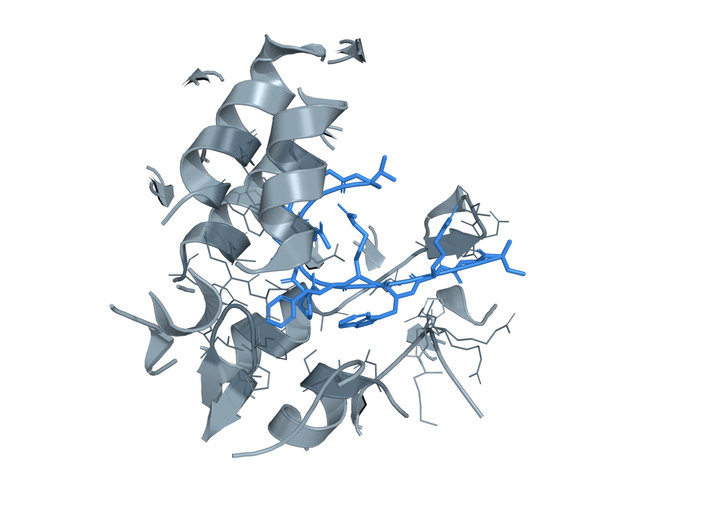
PyMOL render. Menin's pocket (grey; cartoon and lining side chains within 12 Å of the peptide) with KMT2A residues 4-15 (blue), the menin-binding motif every KMT2A fusion retains.
4GQ6 The interface the whole therapeutic programme is aimed at: 12 residues of KMT2A (MBM1) buried in menin's central cavity. This peptide is retained intact by the fusion.
Coordinates stream live from RCSB; nothing here is a drawing of a structure. Domain boundaries are the curated values in gene-kmt2a and protein-aff1-domains. Method and resolution for all eleven entries were read from the RCSB REST API rather than from memory. The junction itself has never been solved, and the residues either side of it are predicted to be disordered — which is the honest reason there is no picture of this protein.
- ACIN1::NUTM1 - the commonest NUTM1 fusion in infants, cryptic to cytogenetics, and the only one with a human cell line · bp-acin1-exon-3-nutm1-exon-3 — Isoform 1 of two. The founding report identifies ACIN1 exon 3 OR exon 4 joined to NUTM1 exon 3, both retaining the functional DNA-binding SAP domain and most of NUTM1 (PMID 31515871). frequency_pct is null because neither isoform's share is reported. The domains_lost_5 list follows from the SAP domain sitting at aa 72-106 while everything else annotated in ACIN1 is C-terminal of residue 1000 (UniProt Q9UKV3, fetched live); it is an inference from domain position, not a statement the source makes.
- ACIN1::NUTM1 - the commonest NUTM1 fusion in infants, cryptic to cytogenetics, and the only one with a human cell line · bp-acin1-exon-4-nutm1-exon-3 — Isoform 2 of two, from the same founding report. No functional difference between the two isoforms is reported.
- BCR::ABL1 · bp-bcr-e13e14-abl1-exon2-p210 — The p210 chimera; defines chronic myeloid leukaemia. Higher kinase activity and transformation potency than p190 in matched assays [E2]. The vacated myristate pocket is the binding site later exploited by the allosteric inhibitor asciminib.
- BCR::ABL1 · bp-bcr-e1-abl1-exon2-p190 — The p190 chimera; predominates in Ph+ ALL, especially paediatric. Lower kinase output than p210 but still fully transforming [E2]. The frequency_pct is the approximate share of transcript types within paediatric Ph+ ALL, not a share of all ALL; the p190/ALL mapping is a strong association rather than a rule.
- BCR::ABL1 · bp-bcr-e19-abl1-exon2-p230 — The mu-bcr p230 chimera, classically associated with a neutrophilic, thrombocytosis-predominant indolent phenotype. Rare; frequency left null because we did not find a cohort-level percentage we could verify.
- BMP2K::ZNF384 - a PROTEIN KINASE as the 5' partner, with its catalytic domain retained: the counterexample to 'the partner is always a chromatin coactivator' · bp-bmp2k-exon15-znf384-exon3 — the case was part of the ZNF384-fusion group sharing weak CD10 with aberrant CD13 and/or CD33; no partner-specific phenotype was reported
- BRD9::NUTM1, t · bp-brd9-exon-variable-nutm1 — BRD9 breakpoints are reported at exons 3, 8 AND 14 in a 9-patient series (PMID 40000924); this record carries the earliest of the three as gene5_exon_last and the other two are listed in data.brd9_breakpoint_exons. NO source read here maps those exons onto BRD9's bromodomain at aa 136-240, so whether the bromodomain is retained is UNKNOWN and is deliberately not asserted. frequency_pct is null because the 9-patient series does not report the per-exon split. The NUTM1 side retains almost the entire open reading frame in every reported NUTM1 fusion, which is why the TADs at 403-470 are listed as retained.
- CBFA2T3::GLIS2, inv · bp-cbfa2t3-nhr-glis2-zinc-fingers — Partner-side blocks are shown in order at nominal width; no residue coordinates are recorded for them.
- CBFB::MYH11 · bp-cbfb-aa165-myh11-coiled-coil — The junction joining the first 165 amino acids of CBFbeta to the SMMHC tail is the one demonstrated in six of six patient samples [E1]. Multiple junction types (conventionally A through J) have been described in the literature, with type A predominating; we state 85% for type A as an approximation of that predominance and flag it as unverified. Exon numbers are convention labels; the reliable statement is the amino-acid boundary (first 165 residues of CBFB) from [E1].
- DUX4::IGH · bp-d4z4-dux4-insertion-igh — Partner-side blocks are shown in order at nominal width; no residue coordinates are recorded for them.
- DUX4::IGH · bp-d4z4-dux4-insertion-erg — Partner-side blocks are shown in order at nominal width; no residue coordinates are recorded for them.
- EP300::ZNF384 - the commonest ZNF384 fusion, and it throws away the acetyltransferase · bp-ep300-exon6-znf384-exon3 — dull or negative CD10, negative cytoplasmic mu chain, aberrant CD13 and/or CD33 in all six founding cases; older median age (11 years) than TCF3::ZNF384
Read this as a table instead
| Break | Share of cases | Kept | Lost |
|---|---|---|---|
| ACIN1::NUTM1 - the commonest NUTM1 fusion in infants, cryptic to cytogenetics, and the only one with a human cell line · bp-acin1-exon-3-nutm1-exon-3 | — | SAP (SAF-A/B, Acinus and PIAS), TAD1 (transactivation domain 1) | — |
| ACIN1::NUTM1 - the commonest NUTM1 fusion in infants, cryptic to cytogenetics, and the only one with a human cell line · bp-acin1-exon-4-nutm1-exon-3 | — | SAP (SAF-A/B, Acinus and PIAS), TAD1 (transactivation domain 1) | — |
| BCR::ABL1 · bp-bcr-e13e14-abl1-exon2-p210 | 95% | OD (oligomerisation domain, structural), Ser/Thr kinase region, ABL SH2-binding region, DH (Dbl homology, RhoGEF), SH3, SH2, PK (tyrosine kinase domain), DNA-binding region | Rho-GAP |
| BCR::ABL1 · bp-bcr-e1-abl1-exon2-p190 | 70% | OD (oligomerisation domain, structural), SH3, SH2, PK (tyrosine kinase domain), DNA-binding region | Ser/Thr kinase region, ABL SH2-binding region, DH (Dbl homology, RhoGEF), Rho-GAP |
| BCR::ABL1 · bp-bcr-e19-abl1-exon2-p230 | — | OD (oligomerisation domain, structural), Ser/Thr kinase region, DH (Dbl homology, RhoGEF), SH3, SH2, PK (tyrosine kinase domain), DNA-binding region | — |
| BMP2K::ZNF384 - a PROTEIN KINASE as the 5' partner, with its catalytic domain retained: the counterexample to 'the partner is always a chromatin coactivator' · bp-bmp2k-exon15-znf384-exon3 | — | PK (protein kinase), ZF1 (C2H2 zinc finger) | — |
| BRD9::NUTM1, t · bp-brd9-exon-variable-nutm1 | — | TAD1 (transactivation domain 1) | — |
| CBFA2T3::GLIS2, inv · bp-cbfa2t3-nhr-glis2-zinc-fingers | 100% | TAFH, Nervy homology region 2 (NHR2); essential for down-regulation of PFKFB3, PFKFB4 and PDK1 expression, Nervy homology region 3 (NHR3); essential for down-regulation of PFKFB3, PFKFB4 and PDK1 expression, C2H2-type 1, GLIS2 C-terminus | MYND-type |
| CBFB::MYH11 · bp-cbfb-aa165-myh11-coiled-coil | 85% | CBFB core (Runt-binding beta-barrel fold), CC (coiled-coil rod, multimerisation) | CBFB core (Runt-binding beta-barrel fold) |
| DUX4::IGH · bp-d4z4-dux4-insertion-igh | 85% | Homeobox 1, IGH Emu enhancer, IGH 3-prime regulatory region enhancers - regulatory elements; no annotated coding exon, though junction read-through into IGH-locus sequence is translated onto DUX4's C terminus | Homeobox 1 |
| DUX4::IGH · bp-d4z4-dux4-insertion-erg | 15% | Homeobox 1, ERG locus regulatory sequence - recipient environment only | Homeobox 1 |
| EP300::ZNF384 - the commonest ZNF384 fusion, and it throws away the acetyltransferase · bp-ep300-exon6-znf384-exon3 | 89% | TAZ1 (TAZ-type zinc finger 1), ZF1 (C2H2 zinc finger) | KIX, BRD (bromodomain), TAZ1 (TAZ-type zinc finger 1) |
▸How exact are these residue numbers?
Domain boundaries are approximate and drawn from UniProt annotation and the cited structural literature; residue ranges for the PHD/bromo region in particular vary by a few residues between sources. Treat the ruler as a schematic of order and extent, not as exact coordinates.
| Fusion | 5′ gene | 3′ gene | Cytoband | Diseases | Breaks | Sources |
|---|---|---|---|---|---|---|
| ACIN1::NUTM1 - the commonest NUTM1 fusion in infants, cryptic to cytogenetics, and the only one with a human cell line | ACIN1 | NUTM1 | t(14;15)(q11.2;q14) | infant B-cell precursor ALL (KMT2A-wild-type), paediatric B-ALL | 2 | 6 |
| AKAP8::ZNF384 - the same protein that MLL-rearranged leukaemia depends on is itself a rare B-ALL fusion partner, on the 5' side, donating its condensate-forming IDR | AKAP8 | ZNF384 | AKAP8 19p13.12; ZNF384 12p13.31 | B-cell precursor acute lymphoblastic leukaemia (adult B-other) | 0 | 3 |
| BCR::ABL1 (Philadelphia chromosome), t(9;22)(q34;q11) | BCR | ABL1 | t(9;22)(q34;q11) | chronic myeloid leukemia, pediatric Ph-positive B-cell precursor ALL, adult Ph-positive B-ALL, Ph-positive AML (rare), CML blast crisis | 3 | 11 |
| BMP2K::ZNF384 - a PROTEIN KINASE as the 5' partner, with its catalytic domain retained: the counterexample to 'the partner is always a chromatin coactivator' | BMP2K | ZNF384 | BMP2K 4q21.21; no karyotype reported for this case in what was read | childhood B-cell precursor ALL | 1 | 3 |
| BRD9::NUTM1, t(5;15)(p15.33;q14) - one fusion is enough to make infant B-ALL in a mouse, and it is the one that behaves best in a child | BRD9 | NUTM1 | t(5;15)(p15.33;q14) | infant B-cell precursor ALL (KMT2A-wild-type), paediatric B-ALL | 1 | 7 |
| CBFA2T3::GLIS2, inv(16)(p13.3q24.3) | CBFA2T3 | GLIS2 | inv(16)(p13.3q24.3) | pediatric non-Down-syndrome acute megakaryoblastic leukemia (AMKL, FAB M7), pediatric non-megakaryoblastic AML (minority) | 1 | 7 |
| CBFB::MYH11 (CBFbeta-SMMHC), inv(16)(p13;q22) | CBFB | MYH11 | inv(16)(p13;q22) | AML FAB M4Eo (acute myelomonocytic leukemia with abnormal eosinophils), pediatric AML, adult AML, AML with myeloid sarcoma, AML with CNS involvement | 1 | 7 |
| CCDC32::CBX3, a previously unreported second fusion found alongside KMT2A::AFDN in a relapsing AML - and the only record in PubMed that pairs CBX3 with KMT2A at all | CCDC32 | CBX3 | CCDC32 15q15.1 :: CBX3 7p15.2 | KMT2A::AFDN-rearranged acute myeloid leukaemia | 0 | 2 |
| DUX4::IGH (IGH-DUX4), t(4;14)/ins(14;4) — D4Z4 repeat insertion into the IGH locus | DUX4 | IGH | ins(14;4)(q32;q35) / t(4;14)(q35;q32) - insertion of D4Z4/DUX4 from 4q35 into the IGH locus at 14q32 | B-cell precursor ALL with DUX4 rearrangement (WHO/ICC entity), adolescent and young adult B-ALL, pediatric B-ALL, adult B-ALL | 2 | 8 |
| EP300::ZNF384 - the commonest ZNF384 fusion, and it throws away the acetyltransferase | EP300 | ZNF384 | t(12;22)(p13;q13) - CRYPTIC: not detected by conventional G-banding in any of the six founding cases, five of which had a normal karyotype | childhood and adult B-cell precursor ALL, B/myeloid MPAL | 1 | 6 |
| ETV6::NTRK3 (ETV6-TRKC), t(12;15)(p13;q25) | ETV6 | NTRK3 | t(12;15)(p13;q25) | congenital/infantile fibrosarcoma, cellular mesoblastic nephroma, secretory breast carcinoma, secretory carcinoma of salivary gland (mammary analogue), rare NTRK-fusion AML and thyroid carcinoma | 1 | 6 |
| ETV6::RUNX1 (TEL-AML1), t(12;21)(p13;q22) | ETV6 | RUNX1 | t(12;21)(p13;q22) | pediatric B-cell precursor ALL, adolescent B-ALL (uncommon), adult B-ALL (rare) | 1 | 12 |
| EWSR1::FLI1 (EWS-FLI1), t(11;22)(q24;q12) | EWSR1 | FLI1 | t(11;22)(q24;q12) | Ewing sarcoma of bone, extraskeletal Ewing sarcoma, peripheral primitive neuroectodermal tumour | 2 | 6 |
| EWSR1::ZNF384 - the same EWSR1 exon 7 fragment that drives Ewing sarcoma, fused instead to a zinc-finger protein, in B-ALL | EWSR1 | ZNF384 | t(12;22)(p13;q12) | B-cell precursor ALL, acute undifferentiated leukaemia (one founding case) | 2 | 5 |
| KMT2A::ABI1 (MLL-ABI1), t(10;11)(p11.2;q23) — the partner that binds an elongation partner | KMT2A | ABI1 | t(10;11)(p11.2;q23) | infant AML, paediatric AML, congenital monocytic leukaemia | 1 | 8 |
| KMT2A::AFDN (MLL-AF6), t(6;11)(q27;q23) | KMT2A | AFDN | t(6;11)(q27;q23) | paediatric AML, adult AML, T-ALL (minor-BCR cases), B-ALL (rare) | 2 | 6 |
| KMT2A::AFF1 (MLL-AF4), t(4;11)(q21;q23) | KMT2A | AFF1 | t(4;11)(q21;q23) | infant B-ALL, childhood B-ALL, adult B-ALL, MPAL, t-AML | 2 | 8 |
| KMT2A::ELL (MLL-ELL), t(11;19)(q23;p13.1) | KMT2A | ELL | t(11;19)(q23;p13.1) | paediatric AML, infant AML, adult AML, therapy-related AML | 2 | 9 |
| KMT2A::EPS15 (MLL-AF1p), t(1;11)(p32;q23) — the infant-ALL cytoplasmic partner | KMT2A | EPS15 | t(1;11)(p32;q23) | infant B-ALL, paediatric B-ALL, adult B-ALL, infant AML, paediatric AML, adult AML, acute monocytic leukaemia, therapy-related ALL | 3 | 11 |
| KMT2A::GAS7 (MLL-GAS7), t(11;17)(q23;p13) — three patients, and the best mouse model in the partner class | KMT2A | GAS7 | t(11;17)(q23;p13); recorded in the recombinome as t(11;17)(q23.3;p13.1). Frequently cryptic: the index case presented as del(11)(q23) on karyotype. | therapy-related AML (post topoisomerase-II inhibitor), de novo paediatric precursor-B ALL, infant AML | 1 | 7 |
| KMT2A::GPHN (MLL-gephyrin), t(11;14)(q23;q24) — the 15-residue counterexample | KMT2A | GPHN | t(11;14)(q23;q24) | de novo acute monoblastic leukaemia (AML M5), therapy-related acute undifferentiated leukaemia (paediatric, post-VP16) | 2 | 6 |
| KMT2A::MAML2, inv(11)(q21q23) - a therapy-related fusion that also causes a solid tumour | KMT2A | MAML2 | inv(11)(q21q23) | therapy-related secondary AML, therapy-related MDS, therapy-related T-ALL (paediatric), T-lymphoblastic lymphoma (secondary), B-ALL (one adult case, post-sarcoma), type B2/B3 thymoma | 1 | 10 |
| KMT2A::MAPRE1 (MLL-EB1) - a coiled-coil dimerisation fusion from pro-B ALL with a mouse model, low leukaemogenic potential, and Ets1 as its effector | KMT2A | MAPRE1 | — | — | 0 | 2 |
| KMT2A::MLLT1 (MLL-ENL), t(11;19)(q23;p13.3) | KMT2A | MLLT1 | t(11;19)(q23;p13.3) | infant B-ALL, childhood B-ALL, T-ALL, AML, MPAL | 2 | 6 |
| KMT2A::MLLT10 (MLL-AF10), t(10;11)(p12;q23) | KMT2A | MLLT10 | t(10;11)(p12;q23) | infant AML, paediatric AML, adult AML, T-ALL (minority), MPAL | 1 | 6 |
| KMT2A::MLLT11 (MLL-AF1q), t(1;11)(q21;q23) — 28 patients, and not one functional experiment | KMT2A | MLLT11 | t(1;11)(q21;q23); also reported as complex three-way rearrangements including t(1;11;4)(q21;q23;p16) and a three-way translocation with MYO18A | infant AML, paediatric AML, adult AML, infant B-ALL (1 case in 28) | 3 | 7 |
| KMT2A::MLLT3 (MLL-AF9), t(9;11)(p21.3;q23.3) | KMT2A | MLLT3 | t(9;11)(p21.3;q23.3) | paediatric AML, infant AML, adult AML, therapy-related AML, KMT2A-r ALL (minority), MPAL | 2 | 6 |
| KMT2A::MLLT6 (MLL-AF17), t(11;17)(q23;q12-q21) — the bridge case: a leucine zipper on a DOT1L-complex subunit | KMT2A | MLLT6 | t(11;17)(q23;q21) in the founding report and most case reports, written t(11;17)(q23;q12-q21) in several; the 2023 recombinome's partner table places MLLT6 at 17q12 and writes t(11;17)(q23.3;q12). The discrepancy is recorded, not resolved. | adult AML, paediatric AML, infant B-ALL, paediatric ALL, acute megakaryoblastic leukaemia (one reported case) | 1 | 6 |
| KMT2A::MYO1F, t(11;19)(q23;p13.2) — recurrent in infant AML, and never functionally tested | KMT2A | MYO1F | t(11;19)(q23;p13.2); the index case was a complex translocation involving chromosomes 7, 11, 19 and 22 | infant AML, infant acute monocytic leukaemia, paediatric AML | 1 | 4 |
| KMT2A::SEPTIN11 (MLL-SEPT11) — the fusion that cytogenetically impersonates t(4;11) | KMT2A | SEPTIN11 | t(4;11)(q21;q23) — cytogenetically indistinguishable from KMT2A::AFF1 | adult ALL, paediatric ALL, paediatric AML | 1 | 5 |
| KMT2A::SEPTIN2 (MLL-SEPT2), t(2;11)(q37;q23) — the therapy-related septin | KMT2A | SEPTIN2 | t(2;11)(q37;q23) | therapy-related AML, therapy-related MDS, paediatric AML | 1 | 8 |
| KMT2A::SEPTIN5 (MLL-SEPT5 / MLL-hCDCrel-1), t(11;22)(q23;q11.2) | KMT2A | SEPTIN5 | t(11;22)(q23;q11.2) | infant AML, paediatric AML, adult AML, paediatric ALL (one case), MDS | 1 | 7 |
| KMT2A::SEPTIN6 (MLL-SEPT6), t(X;11)(q24;q23) and its cryptic insertion variants | KMT2A | SEPTIN6 | t(X;11)(q24;q23); frequently cryptic inv ins(X;11) or three-way rearrangements | infant AML, paediatric AML, adult AML | 2 | 8 |
| KMT2A::SEPTIN9 (MLL-SEPT9 / MLL-MSF), t(11;17)(q23;q25) | KMT2A | SEPTIN9 | t(11;17)(q23;q25) | paediatric AML, adult AML, therapy-related AML, de novo MDS, acute megakaryoblastic leukaemia | 1 | 8 |
| KMT2A::TET1 (MLL-LCX), t(10;11)(q22;q23) — the fusion that throws away a second CXXC domain | KMT2A | TET1 | t(10;11)(q22;q23) | — | 0 | 4 |
| KMT2A::USP2, inv(11)(q23.3q23.3) - the minor-BCR fusion that keeps the PHD fingers | KMT2A | USP2 | inv(11)(q23.3q23.3) | paediatric B-ALL, mixed-phenotype acute leukaemia (B/myeloid), infant AML | 1 | 13 |
| KMT2A::USP8, t(11;15)(q23;q21) - three patients, and the second instance of a deubiquitinase partner | KMT2A | USP8 | t(11;15)(q23;q21) | paediatric B-ALL | 1 | 5 |
| LDB1::KMT2A - the clearest statement in this literature that a sarcoma fusion keeps KMT2A exons 4 to 36 and discards exons 1 to 3 | LDB1 | KMT2A | — | KMT2A-rearranged soft tissue sarcoma, spindle-cell | 1 | 5 |
| MBTD1::EZHIP (MBTD1-CXorf67), t(X;17)(p11.2;q21.33) - MBTD1's other fusion, in which a NuA4 subunit delivers an H3K27M-mimicking PRC2 poison to low-grade endometrial stromal sarcoma | MBTD1 | EZHIP | t(X;17)(p11.2;q21.33), reciprocal | low-grade endometrial stromal sarcoma, classical histology | 1 | 13 |
| NPM1::ALK (NPM-ALK), t(2;5)(p23;q35) | NPM1 | ALK | t(2;5)(p23;q35) | ALK-positive anaplastic large-cell lymphoma, ALK-positive diffuse large B-cell lymphoma (rare) | 1 | 6 |
| NUP98::DDX10, inv(11)(p15q22) — the 'helicase' fusion whose helicase activity turns out not to be the point | NUP98 | DDX10 | inv(11)(p15q22) | de novo AML, therapy-related AML, MDS | 1 | 5 |
| NUP98::HOXA9, t(7;11)(p15;p15) — the founding NUP98 fusion, and an incomplete oncogene | NUP98 | HOXA9 | t(7;11)(p15;p15) | AML (FAB M2, M4), myeloproliferative disease in mouse models | 1 | 10 |
| NUP98::HOXD13, t(2;11)(q31;p15) — the myelodysplasia fusion, and a warning against targeting p53 | NUP98 | HOXD13 | t(2;11)(q31;p15) | therapy-related AML, de novo infant acute myelomonocytic leukaemia, MDS, CML blast crisis | 1 | 8 |
| NUP98::KDM5A (NUP98-JARID1A), t(11;12)(p15;p13) | NUP98 | KDM5A | t(11;12)(p15;p13) | pediatric non-Down-syndrome acute megakaryoblastic leukemia (AMKL, FAB M7), pediatric AML (non-megakaryoblastic, minority) | 1 | 7 |
| NUP98::KMT2A, inv(11)(p15q23) — the NUP98 fusion that contains KMT2A and is menin-inhibitor RESISTANT | NUP98 | KMT2A | inv(11)(p15q23) | AML, MDS | 1 | 4 |
| NUP98::NSD1, t(5;11)(q35;p15.5) | NUP98 | NSD1 | t(5;11)(q35;p15.5) | pediatric de novo AML (FAB M4/M5 predominant), adult AML, therapy-related AML, MDS (rare) | 1 | 8 |
| NUP98::TOP1, t(11;20)(p15;q11~q12) — the most precisely mapped NUP98 breakpoint, and the reciprocal that was wrong twice | NUP98 | TOP1 | t(11;20)(p15;q11~q12) | therapy-related MDS, de novo AML (FAB M2) | 1 | 6 |
| PICALM::MLLT10 (CALM-AF10), t(10;11)(p13;q14) - the DOT1L-dependent HOXA fusion that contains no KMT2A | PICALM | MLLT10 | t(10;11)(p13;q14-21) | T-ALL (including ETP-ALL), AML, acute leukaemia of ambiguous lineage / MPAL, rarely B-ALL | 1 | 17 |
| PML::RARA, t(15;17)(q24;q21) | PML | RARA | t(15;17)(q24;q21) | acute promyelocytic leukemia (APL, FAB M3 and M3v microgranular variant), pediatric APL, adult APL | 2 | 8 |
| RUNX1::RUNX1T1 (AML1-ETO), t(8;21)(q22;q22) | RUNX1 | RUNX1T1 | t(8;21)(q22;q22) | pediatric AML (FAB M2 predominant), adult AML, AML with myeloid sarcoma / extramedullary disease, rare FAB M0, M1, M4 | 2 | 7 |
| SET::NUP214 (TAF-Ibeta-NUP214, SET-CAN), cryptic del(9)(q34.11q34.13) - a HOXA-driving fusion of a histone chaperone to a nucleoporin, with no KMT2A and no menin-binding motif | SET | NUP214 | del(9)(q34.11q34.13); also reported as balanced t(9;9)(q34;q34) | adult T-ALL, paediatric T-ALL, AML, acute undifferentiated leukaemia, acute megakaryoblastic leukaemia | 1 | 9 |
| TAF15::ZNF384 - the FET-partner version, where the naming order in the literature is a trap and the founding experiment says the transactivation domain is not doing the work | TAF15 | ZNF384 | t(12;17) - the 17q band is written q11, q12 or q21 across six papers; TAF15 maps to 17q12 | childhood and adult B-cell precursor ALL, childhood mixed-phenotype (B/myeloid) acute leukaemia, acute undifferentiated leukaemia and AML-M1 in the founding series | 2 | 5 |
| TCF3::HLF (E2A-HLF), t(17;19)(q22;p13) | TCF3 | HLF | t(17;19)(q22;p13) | pediatric pro-B / early B-cell precursor ALL, adolescent B-ALL | 1 | 8 |
| TCF3::PBX1 (E2A-PBX1), t(1;19)(q23;p13.3) | TCF3 | PBX1 | t(1;19)(q23;p13.3) | pediatric B-cell precursor ALL (pre-B, cytoplasmic mu positive), adolescent B-ALL, adult B-ALL (rare) | 1 | 6 |
| TCF3::ZNF384 - the partner whose contribution is now actually demonstrated: steroid resistance, and it is not shared with EP300::ZNF384 | TCF3 | ZNF384 | t(12;19)(p13;p13) - reported as a cryptic t(12;19)(p13.3;p13.3) in the earliest description of E2A-ZNF384 | childhood B-cell precursor ALL, B/myeloid MPAL | 2 | 6 |
| VIM::KMT2A - the one KMT2A rearrangement that keeps the SET domain and still throws away the menin-binding exon | VIM | KMT2A | VIM 10p13 / KMT2A 11q23.3 (interchromosomal, unlike the intrachromosomal YAP1 arm) | KMT2A-rearranged soft tissue sarcoma, spindle-to-round cell subtype | 1 | 6 |
| YAP1::KMT2A::YAP1 (YKY) - an insertional KMT2A rearrangement that keeps the CXXC zinc finger and throws the menin-binding exon away | YAP1 | KMT2A | intrachromosomal 11q22.1-22.2 / 11q23.3; the two genes lie ~16 Mb apart on the same strand and in the same transcriptional direction | KMT2A-rearranged soft tissue sarcoma, MUC4-negative sclerosing epithelioid fibrosarcoma | 2 | 6 |
| ZMYND11::MBTD1, t(10;17)(p15;q21) - a chromatin reader welded to the NuA4/TIP60 acetyltransferase, maintaining Hoxa/Meis1/Myb/Myc/Sox4 in AML: the KMT2A-fusion architecture built from different parts | ZMYND11 | MBTD1 | t(10;17)(p15;q21); refined by optical genome mapping to t(10;17)(p15.3;q21.33) | acute myeloid leukaemia with minimal differentiation (FAB M0), myeloid/NK cell precursor leukaemia (proposed reclassification of the same immunophenotype), therapy-related AML (one reported case) | 2 | 11 |
| ZNF384::TEX41 - the case that breaks the family's architecture: ZNF384 is the 5' partner, the 3' partner is a lncRNA, and the fusion is out of frame | ZNF384 | TEX41 | ZNF384 12p13 to TEX41 2q22.3; the paper reports the fusion from RNA-seq and this entry records no karyotype | childhood B-other BCP-ALL with the ZNF384-rearranged expression signature | 1 | 3 |
Where in the gene the cut lands, and what that costs the protein.
Each partner prefers a different KMT2A exon: partner-specific break position in 3,401 patients
The place where KMT2A snaps is not random, and it is not the same for every partner: AFF1 fusions in babies usually break at one spot while AFDN fusions usually break at another. That means the partner and the break position arrive together, so you can never ask what the break position does without also asking which partner came with it.
KMT2A exon 11 / intron 11 breakpoints (BCR subregion C): the infant, HOXA-low, topoisomerase-II-associated break
The second hot spot for KMT2A breaks sits in intron 11, a piece of DNA that is unusually easy to cut. Babies under six months and patients whose leukaemia was caused by earlier chemotherapy break here most often, and this is the break linked to worse survival.
KMT2A intron 9 / exon 9 breakpoints (BCR subregion A): the HOXA-high, older-patient break
One of the two hot spots inside the KMT2A break region sits in intron 9, near the front of the gene. Leukaemias that break here tend to arise in older children and adults and to switch on the HOXA gene cluster strongly.
KMT2A major breakpoint cluster region (BCR1): intron 7 - exon 13, ~8.3 kb
Almost every leukaemia-causing break in the KMT2A gene happens inside one small stretch of DNA about 8,300 letters long. Where inside that stretch the break lands decides which working parts of the KMT2A protein survive into the fusion protein.
KMT2A minor breakpoint cluster region (BCR2): intron 20 - exon 24
A small minority of KMT2A breaks (about 1 in 70) happen much further along the gene, past the chromatin-reading fingers. The fusion proteins these make are a different beast: they keep parts that every common KMT2A fusion throws away.
MAML2 exon 2: the strongly preferred 3' acceptor, the same in leukaemia, thymoma and salivary-gland tumours
When MAML2 is joined to another gene in a tumour, the join is almost always at the same place - the start of its second exon - no matter which gene it is joined to or which organ the tumour is in. One published patient is the exception, so this is a strong preference rather than a rule, and that kind of near-sameness usually means the piece that survives is the piece that matters.
Spliced KMT2A fusions join to partner exon 2 — with the per-partner counts, and a correction
In some cases the break lands in front of the partner gene rather than inside it, and the cell then splices KMT2A's last surviving exon straight onto the partner's second exon. This entry lists how often that happens for each partner, and corrects an existing entry in this graph that attributed the phenomenon to the wrong gene.
Spliced KMT2A fusions: breaks upstream of the partner's exon 1, and the reciprocal-free natural experiment
Sometimes the break does not land inside the partner gene at all - it lands in front of it. The KMT2A promoter then simply reads straight through into the intact partner gene, and the cell splices the two together. These cases make only one hybrid protein instead of two.
The "second breakpoint cluster region" of KMT2A (Meyer 2019): the founding report of BCR2, not a third cluster
A 2019 paper titled "a second breakpoint cluster region" is easily read as announcing a brand-new, third place where the KMT2A gene breaks. It is not: it is the paper that first found the region this graph already calls the minor BCR, and reading the title without the paper would count one region twice.
The KMT2A breakpoint cluster region: an 8.3 kb stretch built to break
Almost every KMT2A fusion snaps inside the same short 8.3 kilobase stretch of the gene, which happens to be loaded with features that make DNA easy to cut: places where the DNA is unwound and exposed, anchors that tether it to the nuclear scaffold, and preferred cutting sites for the enzyme topoisomerase II. The gene is not simply unlucky, that piece of it is structurally primed to break.
Where t(9;11) actually breaks inside MLLT3: 98.4% downstream of exon 4, and the two patients in 442 who kept the reader
In 442 patients with this translocation, the chromosome broke past AF9's fourth exon in all but a handful, throwing the reader module away every time. Exactly two patients kept it, and nothing at all is known about those two.
Where the break falls, and the three incompatible numbering frames that make it easy to get wrong
The chromosome break can fall in several places, producing at least eight different versions of the fusion, and the versions behave differently. Worse, different papers count the amino acids in different ways, so the same position has three different numbers in the literature.
Which partner, in which patient: KMT2A partner frequencies across six age-lineage strata
Which gene KMT2A gets stuck to depends strongly on how old the patient is and whether the leukemia is lymphoid or myeloid. This entry gives the exact patient counts for all six combinations, so that a figure can weight each fusion by how many children it actually affects.
Each partner gene and the disease it tends to produce.
ACIN1::NUTM1 - the commonest NUTM1 fusion in infants, cryptic to cytogenetics, and the only one with a human cell line
This is the most common version of the NUTM1-rearranged leukaemia that affects babies without a KMT2A rearrangement. It joins the DNA-gripping front end of a splicing protein to almost all of NUTM1, and it is invisible to the standard chromosome test - it was found only when researchers sequenced the RNA.
AKAP8::ZNF384 - the same protein that MLL-rearranged leukaemia depends on is itself a rare B-ALL fusion partner, on the 5' side, donating its condensate-forming IDR
AKAP95 turns up twice in leukaemia, in two completely different ways. In MLL-rearranged leukaemia it is a helper the cancer needs. In a rare kind of B-ALL it is itself half of the fusion gene that causes the disease.
BCR::ABL1 (Philadelphia chromosome), t(9;22)(q34;q11)
A swap between chromosomes 9 and 22 welds a self-clumping piece of one protein onto an enzyme that normally tells blood cells to divide, and the weld jams the enzyme permanently on. Because that enzyme has a real pocket where a drug can sit, a pill called imatinib turned a fatal leukaemia into a manageable condition.
BMP2K::ZNF384 - a PROTEIN KINASE as the 5' partner, with its catalytic domain retained: the counterexample to 'the partner is always a chromatin coactivator'
Almost every description of this leukaemia family says the front half is a chromatin regulator. In this case the front half is an enzyme that adds phosphate groups to other proteins, and the fusion keeps the enzyme part. One patient, but it is enough to show the rule is not a rule.
BRD9::NUTM1, t(5;15)(p15.33;q14) - one fusion is enough to make infant B-ALL in a mouse, and it is the one that behaves best in a child
This is a chromosome accident that joins part of BRD9, a chromatin-reading protein, to almost all of NUTM1, a protein that switches on a histone-modifying enzyme. It causes leukaemia in babies who do not have the more familiar KMT2A rearrangement - and unlike that one, children with this fusion nearly always survive standard chemotherapy.
CBFA2T3::GLIS2, inv(16)(p13.3q24.3)
In some infants and toddlers, a single chromosome folds back on itself and joins a gene that normally silences blood genes to a gene that normally relays a developmental signal, and the hybrid switches on a growth signal that makes immature platelet-precursor cells copy themselves endlessly. It is one of the most aggressive childhood leukaemias, it has a distinctive fingerprint on the flow cytometer, and it has produced the most promising immune target in this whole family of leukaemias.
CBFB::MYH11 (CBFbeta-SMMHC), inv(16)(p13;q22)
One chromosome folds back on itself and joins the helper half of a master blood-cell switch to the long rope-like tail of a muscle protein. The hybrid grabs the switch's business half more tightly than the real helper does, drags it away from the genes it should be turning on, and produces a leukaemia with a distinctive abnormal eosinophil - and, unusually, a drug has been built that pries the two apart.
CCDC32::CBX3, a previously unreported second fusion found alongside KMT2A::AFDN in a relapsing AML - and the only record in PubMed that pairs CBX3 with KMT2A at all
In a young adult whose leukaemia kept coming back, sequencing found a second, previously unknown gene fusion sitting alongside the KMT2A fusion that caused the disease. The second fusion involves CBX3, a protein this project already tracks for a different reason, and it changed the activity of both genes it is made from.
DUX4::IGH (IGH-DUX4), t(4;14)/ins(14;4) — D4Z4 repeat insertion into the IGH locus
A gene that is normally switched off in every cell except the early embryo and testis gets physically dropped into the antibody gene factory of a young B cell, where the cell's most powerful gene switch is permanently on. That switch drives a truncated version of the embryonic gene, which then wrecks a second gene called ERG - and, unusually for such a strange accident, children with this leukaemia do well.
EP300::ZNF384 - the commonest ZNF384 fusion, and it throws away the acetyltransferase
This is the most frequent version of the ZNF384 fusion. The partner, EP300, is a famous enzyme that tags chromatin with acetyl marks - and the fusion keeps only the first fifth of it and discards the enzyme part entirely. Children with this version do better than those with the other common versions.
ETV6::NTRK3 (ETV6-TRKC), t(12;15)(p13;q25)
A gene called ETV6, whose job includes sticking to copies of itself, is fused to the enzyme half of a nerve-growth-factor receptor called TRKC, so the enzyme is forced into a long chain of active copies and never switches off. Because that enzyme has a real ATP pocket, a pill can shut it down - and in babies with congenital fibrosarcoma this has replaced disfiguring surgery.
ETV6::RUNX1 (TEL-AML1), t(12;21)(p13;q22)
In about a quarter of children with the commonest childhood leukaemia, two chromosomes swap ends so that a gene that normally silences genes gets bolted onto a gene that normally switches blood genes on, and the hybrid silences the genes it should activate. The first half of this accident usually happens before birth, and most children who then develop the leukaemia are cured.
EWSR1::FLI1 (EWS-FLI1), t(11;22)(q24;q12)
In Ewing sarcoma, a bone and soft-tissue cancer of teenagers, half of a gene called EWSR1 - a floppy, sticky, low-complexity stretch of protein - is fused to the DNA-gripping end of FLI1. The sticky half recruits chromatin machinery to short repeated DNA sequences that are normally silent junk, switching on hundreds of genes that should never be on. There is no pocket anywhere on this protein, which is why after thirty years there is still no targeted drug.
EWSR1::ZNF384 - the same EWSR1 exon 7 fragment that drives Ewing sarcoma, fused instead to a zinc-finger protein, in B-ALL
The front half of this leukaemia fusion is exactly the same piece of EWSR1 that, welded to a different partner, causes Ewing sarcoma - a bone cancer. Same module, different DNA-binding partner, completely different disease.
KMT2A::ABI1 (MLL-ABI1), t(10;11)(p11.2;q23) — the partner that binds an elongation partner
KMT2A here joins ABI1, a protein of the cell's actin machinery that normally helps cells move and change shape. What makes it the most interesting partner in this group is that ABI1 physically binds ENL — one of the classic elongation partners — so this fusion may reach the elongation machinery by a completely different route.
KMT2A::AFDN (MLL-AF6), t(6;11)(q27;q23)
This is the odd one out: the broken KMT2A gene joins afadin, a protein that normally lives outside the nucleus holding cells together at their junctions. Afadin contributes nothing but the ability to stick to itself, which forces two copies of the fusion together, and that alone is enough to switch the leukemia program on - the same trick the curable kinase fusions use.
KMT2A::AFF1 (MLL-AF4), t(4;11)(q21;q23)
In this leukemia a gene called KMT2A snaps in half and glues itself to a gene called AFF1, and the hybrid protein that results sits on the genes a blood cell needs to grow up and jams them permanently switched on. It is the commonest cause of leukemia in babies under one year old, and only about half of those babies are cured, because unlike the kinase fusions this protein has no enzyme pocket for a drug to plug.
KMT2A::ELL (MLL-ELL), t(11;19)(q23;p13.1)
Here the broken KMT2A gene joins ELL, a genuine component of the machinery that makes RNA polymerase stop pausing and run. But when researchers cut ELL apart, the speed-up part turned out not to be what causes the leukemia - the part that grabs a partner protein called EAF1 is. It is mostly a myeloid leukemia partner, notably common in infant myeloid leukemia and in leukemias caused by previous chemotherapy.
KMT2A::EPS15 (MLL-AF1p), t(1;11)(p32;q23) — the infant-ALL cytoplasmic partner
This fusion joins the leukaemia gene KMT2A to EPS15, a protein whose day job is helping cells swallow things from their surface — nothing to do with reading DNA. It is the eighth most common KMT2A partner, it hits infants with B-cell leukaemia hardest, and it is one of the two cases where a short self-sticking stretch of the partner was shown to be both necessary and sufficient to cause the disease.
KMT2A::GAS7 (MLL-GAS7), t(11;17)(q23;p13) — three patients, and the best mouse model in the partner class
Only three patients with this fusion appear in the largest survey ever done, yet it is one of the most studied KMT2A fusions in mice — because it was the one used to show that a cytoplasmic partner causes leukaemia simply by making copies of KMT2A stick together. In mice it produces three different leukaemias, including one that is simultaneously myeloid and lymphoid.
KMT2A::GPHN (MLL-gephyrin), t(11;14)(q23;q24) — the 15-residue counterexample
This is one of the rarest leukaemia fusions known — only two patients have ever been described — but it is one of the most important, because the piece of the partner protein that makes it cancerous is just fifteen amino acids long and does nothing to genes. It is the clearest evidence that a KMT2A fusion does not need to hijack the cell's gene-reading machinery to cause leukaemia.
KMT2A::MAML2, inv(11)(q21q23) - a therapy-related fusion that also causes a solid tumour
This fusion is made when a piece of chromosome 11 flips, joining KMT2A to MAML2. It is unusual twice over: it usually appears as a second cancer after chemotherapy for something else, and the very same fusion turns up in thymoma, a tumour of the chest.
KMT2A::MAPRE1 (MLL-EB1) - a coiled-coil dimerisation fusion from pro-B ALL with a mouse model, low leukaemogenic potential, and Ets1 as its effector
In one adult patient with pro-B acute lymphoblastic leukaemia, the KMT2A gene was joined to EB1, a gene for a microtubule-tip protein. The joined protein sits in the nucleus and can turn mouse bone marrow cancerous, but it does so weakly compared with other KMT2A fusions.
KMT2A::MLLT1 (MLL-ENL), t(11;19)(q23;p13.3)
Here the broken KMT2A gene joins ENL, a close cousin of the AF9 partner, and the result is usually a lymphoid leukemia - sometimes of B cells, distinctively often of T cells. Its most surprising feature is that outcome depends almost entirely on the patient's age: infants do badly, while children between one and nine and those with T-cell disease have done remarkably well.
KMT2A::MLLT10 (MLL-AF10), t(10;11)(p12;q23)
In this version the broken KMT2A gene joins AF10, and the only part of AF10 that matters is a short coiled stretch that grabs an enzyme called DOT1L and drags it onto the wrong genes. It is one of the commonest partners in infant and childhood myeloid leukemia and one of the worst, and the same DOT1L-grabbing stretch turns up in a completely different fusion, CALM-AF10, which is strong evidence that this one small module is the whole problem.
KMT2A::MLLT11 (MLL-AF1q), t(1;11)(q21;q23) — 28 patients, and not one functional experiment
This is the tenth most common KMT2A leukaemia fusion, found in 28 patients in the largest survey ever done — almost all of them infants and children with myeloid leukaemia. In more than thirty years since it was discovered, nobody has ever put the fusion protein into a cell and asked whether it causes cancer.
KMT2A::MLLT3 (MLL-AF9), t(9;11)(p21.3;q23.3)
This is the same broken KMT2A gene as in infant leukemia, but joined to a different partner called AF9, and the combination produces myeloid leukemia rather than lymphoid. AF9 carries a small reading head that recognises chemical tags on chromatin and brings in an enzyme called DOT1L, so this version of the fusion has an extra, partly druggable step that the AFF1 version does not.
KMT2A::MLLT6 (MLL-AF17), t(11;17)(q23;q12-q21) — the bridge case: a leucine zipper on a DOT1L-complex subunit
This fusion is the awkward one for tidy classifications. Its partner AF17 carries a short self-sticking coil — the hallmark of the "just make two copies stick together" explanation — but AF17 is also a member of the very machine, the DOT1L complex, that the rival explanation says KMT2A fusions hijack. The same protein belongs to both camps.
KMT2A::MYO1F, t(11;19)(q23;p13.2) — recurrent in infant AML, and never functionally tested
A rare but repeating leukaemia fusion of babies, joining KMT2A to a small motor protein that crawls along the cell's internal cables. The people who found it pointed out that the motor brings neither of the two things every explanation of this disease relies on — and nobody has tested it since.
KMT2A::SEPTIN11 (MLL-SEPT11) — the fusion that cytogenetically impersonates t(4;11)
SEPTIN11 sits on chromosome 4 only a short distance from AFF1, so a KMT2A break that joins it produces a picture under the microscope that is indistinguishable from the commonest and best-known KMT2A leukaemia. The only way to tell them apart is a molecular test.
KMT2A::SEPTIN2 (MLL-SEPT2), t(2;11)(q37;q23) — the therapy-related septin
A rare fusion of KMT2A to septin-2 that has turned up almost only in patients whose leukaemia followed earlier chemotherapy. It is the one septin fusion where somebody measured what happens to the normal copies of both genes, and the answer was that both go down.
KMT2A::SEPTIN5 (MLL-SEPT5 / MLL-hCDCrel-1), t(11;22)(q23;q11.2)
The first septin ever found fused to KMT2A, identified in infant twins with the same leukaemia — strong evidence that the break happened once, before birth, in a shared blood cell. The partner is a cell-division septin that also happens to sit in the stretch of chromosome 22 deleted in DiGeorge syndrome.
KMT2A::SEPTIN6 (MLL-SEPT6), t(X;11)(q24;q23) and its cryptic insertion variants
Here the broken KMT2A gene is joined to septin-6, a cytoskeleton protein that normally helps a cell divide and has nothing to do with reading genes. It is the best-studied of the septin fusions, and in mice it causes only a slow blood disorder unless a second mutation is added.
KMT2A::SEPTIN9 (MLL-SEPT9 / MLL-MSF), t(11;17)(q23;q25)
The second most common septin partner: KMT2A is joined to septin-9, a scaffolding protein of the cell's skeleton. Unlike most KMT2A fusions it shows up in adults and in leukaemias caused by earlier chemotherapy, and it is easy to confuse with a completely different t(11;17) leukaemia that is treated in a completely different way.
KMT2A::TET1 (MLL-LCX), t(10;11)(q22;q23) — the fusion that throws away a second CXXC domain
A rare leukaemia rearrangement joins the KMT2A gene to TET1. What makes it interesting is what gets discarded: TET1 normally carries the same kind of DNA-reading module that KMT2A does, and the join removes it. Two proteins that both read the same chemical mark on DNA are fused, and only one of the two readers survives.
KMT2A::USP2, inv(11)(q23.3q23.3) - the minor-BCR fusion that keeps the PHD fingers
KMT2A::USP2 is made when a piece of chromosome 11 flips over and joins the KMT2A gene to a neighbouring gene about a million letters away. It is the ninth most common KMT2A fusion, it mostly affects babies and toddlers, and ordinary chromosome tests often cannot see it at all.
KMT2A::USP8, t(11;15)(q23;q21) - three patients, and the second instance of a deubiquitinase partner
A very rare leukaemia fusion in which KMT2A joins USP8, an enzyme that removes destruction tags from proteins. Only three patients are known, but they matter because they repeat the pattern seen with the much commoner USP2 fusion.
LDB1::KMT2A - the clearest statement in this literature that a sarcoma fusion keeps KMT2A exons 4 to 36 and discards exons 1 to 3
A single reported sarcoma joined the front of a gene called LDB1 to almost the whole of KMT2A, starting at KMT2A's fourth exon. That means it keeps KMT2A's enzyme end but loses the first three exons, including the one menin holds on to.
MBTD1::EZHIP (MBTD1-CXorf67), t(X;17)(p11.2;q21.33) - MBTD1's other fusion, in which a NuA4 subunit delivers an H3K27M-mimicking PRC2 poison to low-grade endometrial stromal sarcoma
The same gene that forms a leukaemia fusion also forms a fusion in a rare uterine cancer, but the other way round - here it is at the front, and the piece it drags along is a decoy that jams the cell's main gene-silencing enzyme. This is the entry that lets this project compare the two, because the only thing that changes between them is which side of the join the shared gene sits on.
NPM1::ALK (NPM-ALK), t(2;5)(p23;q35)
In this lymphoma a gene for a busy nuclear shuttle protein, NPM1, is fused to the business end of a receptor enzyme called ALK, and because NPM1 naturally clumps together it forces the enzyme to switch itself on permanently. That enzyme has a real pocket where ATP normally binds, so a pill that fills the pocket shuts the cancer off - which is why this fusion is largely treatable and KMT2A::AFF1 is not.
NUP98::DDX10, inv(11)(p15q22) — the 'helicase' fusion whose helicase activity turns out not to be the point
This NUP98 fusion attaches to an RNA-unwinding enzyme, and for nearly thirty years the unwinding was assumed to be what mattered. It is not: a short 24-amino-acid stretch that grabs a nucleolar protein does the work, and the leukaemia depends on that protein instead.
NUP98::HOXA9, t(7;11)(p15;p15) — the founding NUP98 fusion, and an incomplete oncogene
The first NUP98 fusion ever found, reported by two labs in the same journal issue in 1996. On its own it does not make acute leukaemia: mice first get a slow overgrowth of blood cells and only later, after acquiring more damage, an acute leukaemia.
NUP98::HOXD13, t(2;11)(q31;p15) — the myelodysplasia fusion, and a warning against targeting p53
A rare NUP98 fusion found after chemotherapy for another cancer and also in infants. Mice carrying it develop a severe pre-leukaemic bone-marrow failure that always turns into leukaemia — and the obvious idea of blocking p53 to relieve the failure makes it worse.
NUP98::KDM5A (NUP98-JARID1A), t(11;12)(p15;p13)
A cryptic swap between chromosomes 11 and 15 joins a sticky, shapeless piece of a nuclear-pore protein to a small module that recognises a specific chemical tag on DNA-packaging proteins. The hybrid parks on genes that should be switched off during development and refuses to let go, causing an aggressive leukaemia of platelet-precursor cells in young children.
NUP98::KMT2A, inv(11)(p15q23) — the NUP98 fusion that contains KMT2A and is menin-inhibitor RESISTANT
This rare rearrangement joins the NUP98 protein to almost the whole of KMT2A — the very gene whose fusions menin drugs were built for. It is the one NUP98 fusion those drugs do not work on, because the join deletes the exact 20 amino acids that menin grips.
NUP98::NSD1, t(5;11)(q35;p15.5)
A cryptic swap between chromosomes 5 and 11 joins a sticky, shapeless piece of a nuclear-pore protein to an enzyme that puts a chemical mark on DNA-packaging proteins. The sticky end makes the hybrid clump into droplets on the genome and the enzyme end locks developmental genes permanently on, producing an aggressive childhood leukaemia that conventional chemotherapy usually fails.
NUP98::TOP1, t(11;20)(p15;q11~q12) — the most precisely mapped NUP98 breakpoint, and the reciprocal that was wrong twice
This fusion has the best-defined join of any in the family: the same two exons are stitched together in every reported patient. It was first thought to happen only after chemotherapy and to make only one of its two possible products — both of those beliefs turned out to be wrong.
PICALM::MLLT10 (CALM-AF10), t(10;11)(p13;q14) - the DOT1L-dependent HOXA fusion that contains no KMT2A
A chromosome swap glues the clathrin adaptor PICALM onto the chromatin protein AF10, making a single hybrid protein that switches on the same HOXA growth genes as the KMT2A leukaemias - but without containing any KMT2A at all. It causes an aggressive leukaemia that is usually T-cell, sometimes myeloid, and responds poorly to standard chemotherapy.
PML::RARA, t(15;17)(q24;q21)
A swap between chromosomes 15 and 17 joins a protein that builds little clumps inside the nucleus to the receptor for vitamin A, and the hybrid jams white-blood-cell development at one stage. Because the vitamin A pocket is still there and the clumping part can be poisoned by arsenic, two non-chemotherapy drugs destroy the hybrid protein outright - turning the most lethal leukaemia into the most curable one.
RUNX1::RUNX1T1 (AML1-ETO), t(8;21)(q22;q22)
A swap between chromosomes 8 and 21 takes the part of a master blood-cell switch that finds its target genes and bolts it onto a protein that silences genes and glues four copies of itself together. The result parks a silencing machine on exactly the genes that tell a cell to grow up, so the cells stay immature - but this is one of the better-outcome childhood leukaemias.
SET::NUP214 (TAF-Ibeta-NUP214, SET-CAN), cryptic del(9)(q34.11q34.13) - a HOXA-driving fusion of a histone chaperone to a nucleoporin, with no KMT2A and no menin-binding motif
A small deletion on chromosome 9 glues a histone-handling protein called SET onto a piece of a nuclear pore protein called NUP214, and the resulting hybrid switches on the HOXA genes in T-cell leukaemia. It reaches those genes without containing any part of KMT2A, the gene this project's main drug target works through.
TAF15::ZNF384 - the FET-partner version, where the naming order in the literature is a trap and the founding experiment says the transactivation domain is not doing the work
In this version the front half comes from TAF15, a cousin of the Ewing sarcoma protein EWSR1, and it donates the same kind of floppy sticky region. The paper that discovered it tested whether the fusion switches on ZNF384's usual target genes - and it does not, which nobody has explained in more than twenty years.
TCF3::HLF (E2A-HLF), t(17;19)(q22;p13)
A rare swap between chromosomes 17 and 19 welds the powerful switch-on end of a B-cell master regulator onto a liver protein's DNA-gripping end, and the hybrid turns on a survival programme that stops young B cells from dying when they should. It is the deadliest subtype of childhood B-cell leukaemia, but it turned out to depend heavily on the protein BCL2, which a drug called venetoclax blocks.
TCF3::PBX1 (E2A-PBX1), t(1;19)(q23;p13.3)
A swap between chromosomes 1 and 19 replaces the DNA-reading end of a master B-cell switch with the DNA-reading end of a developmental patterning protein, so the resulting hybrid switches on a completely foreign set of genes at full power. Children with this leukaemia used to do badly and now do well on intensive chemotherapy, but their leukaemia has an unusual habit of coming back in the brain and spinal fluid.
TCF3::ZNF384 - the partner whose contribution is now actually demonstrated: steroid resistance, and it is not shared with EP300::ZNF384
This version of the ZNF384 fusion uses TCF3 as the front half - the same gene that partners PBX1 in a well-known childhood leukaemia. Children with it are younger, respond worse to steroids and relapse more often, and one experiment has now shown that the steroid resistance really does come from the TCF3 half and not from the shared ZNF384 half.
VIM::KMT2A - the one KMT2A rearrangement that keeps the SET domain and still throws away the menin-binding exon
In a handful of spindle-cell sarcomas the front half of the vimentin gene is joined to almost the whole of the KMT2A gene, which is the reverse of what happens in leukaemia. The joint falls just past KMT2A's first exon, so the resulting protein keeps KMT2A's enzyme end but loses the small piece that menin grips - the piece the menin-inhibitor drugs depend on.
YAP1::KMT2A::YAP1 (YKY) - an insertional KMT2A rearrangement that keeps the CXXC zinc finger and throws the menin-binding exon away
In a rare soft-tissue cancer of young adults, a small piece of the KMT2A gene is cut out and pasted into the middle of the YAP1 gene, instead of the usual head-to-tail gene fusion. The piece that moves carries only KMT2A's DNA-gripping CXXC finger, and leaves behind the very first exon - the one that holds the grip site for menin, the protein that menin-inhibitor drugs act on.
ZMYND11::MBTD1, t(10;17)(p15;q21) - a chromatin reader welded to the NuA4/TIP60 acetyltransferase, maintaining Hoxa/Meis1/Myb/Myc/Sox4 in AML: the KMT2A-fusion architecture built from different parts
A rare leukaemia swap joins the front half of a gene that reads a chemical tag on packaged DNA to a gene that plugs into a machine that loosens that packaging, and the joined protein drags the loosening machine to the wrong genes and holds them switched on. The genes it holds on are the same ones a KMT2A fusion holds on - HOXA, MEIS1, MYB, MYC - which makes it the closest thing in another disease to the fusion this project is about.
ZNF384::TEX41 - the case that breaks the family's architecture: ZNF384 is the 5' partner, the 3' partner is a lncRNA, and the fusion is out of frame
Every textbook description of this leukaemia subtype says ZNF384 is the second half of the fusion and gets a powerful new front end. In one child it was the other way round: ZNF384 was the front half, the back half came from a gene that does not make a protein at all, and the join was out of frame - yet the leukaemia looked exactly like the others.
ABI1 (Abl interactor 1 / e3B1) — WAVE-complex cytoskeletal adaptor, KMT2A partner, and an ENL-binding protein
ABI1 is a connector protein in the machinery that builds and steers the cell's actin skeleton. It is a rare KMT2A fusion partner, and unusually it also sticks to ENL — one of the common KMT2A partners — which could let it reach the gene-reading machinery indirectly.
ABL1 — domain architecture and residue coordinates (with the 1a/1b numbering trap)
ABL1 is an enzyme that adds phosphate tags to other proteins, and it normally keeps itself switched off by folding a greasy tail back against its own working face. The leukaemia fusion cuts that tail off, which is why the enzyme never switches off again, and because two rival ways of counting ABL1's residues differ by exactly nineteen, every number here has to say which one it uses.
ACIN1 (Acinus) - a splicing/apoptosis protein whose only domain is the 35-residue module the leukaemic fusion keeps, and whose entire localization record is uncoded
ACIN1 normally works on messenger RNA splicing and helps condense the DNA of a dying cell. A leukaemia-causing accident keeps only its small DNA-gripping tip and throws away everything else it does - so the leukaemia is not caused by ACIN1 doing its job in the wrong place, but by that tip being used as a postal address for a different protein.
ACSL4 - a CRISPR-screen-derived, functionally validated KMT2A-selective vulnerability in myeloid disease, and a gene whose loss normally protects cells from ferroptosis
ACSL4 activates long polyunsaturated fats so the cell can use them, and KMT2A-rearranged myeloid leukaemia needs it much more than other leukaemias do. Turning it off slows those cells and delays the disease in mice - and feeding the cells the fats back rescues them, which is how the authors showed the fat handling is really the reason.
ACTB / beta-actin (P60709) as a SUBJECT, not a loading control - the dirtiest negative control in the pan-MLL-fusion 42, and the one the paper's authors kept
Beta-actin is the protein that builds the cell's internal cables. It is so abundant that biologists routinely use it as a background reference on a gel - which is exactly why finding it stuck to a cancer protein is suspicious. But actin also genuinely works inside the nucleus, where it is a built-in part of the machines that open up DNA, so this one cannot simply be dismissed.
AFDN / AF6 — domain architecture and residue coordinates
AFDN is a cell-junction protein that becomes a KMT2A fusion partner, and unusually for a partner it normally lives outside the nucleus. This entry pins down where its parts sit, and the surprise is that the small module at its very front end — not anything larger — is the piece that makes the fusion cancerous.
AFF1 / AF4 — domain architecture and residue coordinates
AFF1 is the partner protein most often stuck onto the broken KMT2A gene in infant leukemia, and this entry pins down where each of its working parts sits along the protein chain. Most of AFF1 is a floppy, shapeless string, so several of its "domains" are conventions drawn on a diagram rather than things anyone has measured.
AFF1 / AF4 — the 3' half of the fusion, and a target with almost no structural footing
AFF1 is the protein whose tail end gets welded onto KMT2A in the commonest infant leukemia fusion. Despite being half the cancer protein, almost nothing is known about its shape: the entire Protein Data Bank contains one small piece of it.
AFF1-AS1 - the antisense RNA 151 bp from the index fusion partner's TSS, which Ensembl does not recognise and this project's promoter table cannot see
Right next to AFF1 - the gene that gives this project's index fusion its business end - another gene starts 151 letters away and points the other way. It has never been studied by anyone, and this project's measurement table has no row for it, so every measurement of AFF1's on-switch has silently been a measurement of both.
AFF1::KMT2A (AF4-MLL), the der(4) product: what it is made of, residue by residue
When chromosomes 4 and 11 swap arms, two hybrid genes are made, not one. This entry is the second one: a protein that throws away the front of KMT2A - including the piece the menin drug grabs - and keeps everything behind it, including the enzyme end.
AFF2 (FMR2) — the SEC-L2 scaffold, and the AFF-family member with no dependency anywhere
AFF2 is the fourth member of the family of scaffold proteins that build the cell's transcription-speeding machine. Unlike its relatives it is switched off in most cells and no cell line tested needs it, so it is the family member that tells you the family slot can be empty.
AFF3 (LAF4) — the AFF-family scaffold of SEC-L3, and a KMT2A fusion partner in its own right
AFF3 is one of four closely related scaffold proteins, any of which can build a version of the elongation machine the KMT2A fusion hijacks. AFF3 builds a different version from the one AFF1 builds, and those versions switch on different sets of genes.
AFF4 — the textbook SEC scaffold that KMT2A::AFF1 cells need LESS than other leukemias
AFF4 is the long, floppy protein usually drawn at the centre of the super elongation complex, holding the other parts together. But when you delete it, KMT2A-rearranged leukemia cells are slightly better off than other leukemia cells are, which is the opposite of what the textbook picture predicts.
AFG2A / SPATA5 (Q8NB90) - a 55LCC replisome-proteostasis ATPase that co-purifies with KMT2A and menin, and whose CURRENT symbol cannot retrieve its own source paper or any of its function literature
AFG2A is an engine-like protein that clears stuck machinery off DNA while it is being copied, and losing it causes an ultra-rare childhood brain disease with epilepsy, small head size and deafness. A single 2024 experiment put it next to both KMT2A and menin, the two proteins at the centre of this project - but in cervical cancer cells, and the rest of its own complex is nowhere in the interaction record.
AFG2B / SPATA5L1 (Q9BVQ7) - the second 55LCC ATPase, the only read-queue member with an annotated nuclear pool, pan-essential in 95.5% of lines, and the one that touches PSIP1
AFG2B is the partner engine to AFG2A in a four-part machine that clears stuck proteins off DNA as it is copied, and losing it causes recessive deafness or a severe childhood brain disorder. One 2024 experiment placed it next to both KMT2A and LEDGF, two proteins this project cares about - but in cervical cancer cells, and it is the only read-queue member that UniProt even places in the nucleus.
AGGF1 - an angiogenic G-patch protein whose BioID neighbourhood contains ELL, KMT2A, LEDGF, WDR5, KAT7 and RNA polymerase II
AGGF1 is known as a blood-vessel growth factor, but it also carries RNA-binding modules. Its neighbour list inside a leukaemia cell contains ELL, KMT2A, LEDGF, WDR5 and RNA polymerase II itself.
AHDC1 (Gibbin) - an AT-hook chromatin-architecture factor that is already a cofactor of a DIFFERENT sarcoma fusion oncoprotein, and whose KMT2A breakpoint lands in an untranslated exon
AHDC1, also called Gibbin, sits on the DNA at gene switches and keeps their looping architecture in place, and losing it makes nearby genes get chemically silenced. In one patient's leiomyosarcoma KMT2A broke at exon 15 and joined AHDC1 - but at a piece of AHDC1 that is never translated, so this fusion carries no AHDC1 protein at all.
AKAP8 (AKAP95) - the only nuclear A-kinase anchoring protein, and the one that turned out to be required for MLL-rearranged leukaemia
AKAP8, usually called AKAP95, is a nuclear scaffolding protein: it holds other proteins in the right place rather than doing chemistry itself. In 2026 it was shown that leukaemia cells carrying an MLL (KMT2A) fusion need it, while normal blood-forming cells do not.
ALDH1L2 (Q3SY69) - the second-largest fall in the serine/one-carbon panel (-3.40), named nowhere in this graph, zero of 1,258 DepMap lines require it, and a tumour SUPPRESSOR where it has been studied
ALDH1L2 burns off spare one-carbon units inside mitochondria and makes NADPH while doing it. A menin inhibitor switches it off about ten-fold in KMT2A-rearranged leukaemia cells, which nobody had noticed - and losing it raises damaging oxygen radicals, so this is a fall that could be helping the cancer rather than hurting it.
ALK (anaplastic lymphoma kinase) domain architecture
ALK is the 3' partner of NPM1::ALK and the target of crizotinib, alectinib and lorlatinib. This entry lists where each of its functional parts sits along the protein, measured as residue numbers, so the fusion diagrams can draw which parts survive the break and which are thrown away.
ALKBH5 — the second m6A eraser: a cancer-stem-cell self-renewal role elsewhere, one incidental KMT2A record, and a flat dependency row
ALKBH5 is the other enzyme that strips the chemical tag off RNA. It helps cancer stem cells renew themselves in some settings, but almost no cancer cell line depends on it and nothing has been published about it in KMT2A-rearranged disease.
ANP32E - the eviction chaperone: the only protein in this beat whose job is to take a histone variant OUT of chromatin
Every other chaperone here puts histones in. ANP32E takes one particular variant back out, which is the half of turnover that nobody measures and that this project's clock argument silently assumes.
AP2B1 (beta-2-adaptin) - the only one of AFF4's four two-hybrid partners that is not a hub, and the only one with an echo in this graph's endocytosis biology
AP2B1 is part of the machinery that pulls cargo into cells from the cell surface. It was reported as a partner of AFF4 in one large automated yeast screen, and unlike the other three proteins from that screen it does not stick to everything, which makes it the least dismissible of them.
AQR (Aquarius) - a spliceosomal RNA helicase whose loss impairs MLL-rearranged leukaemia stem cells
AQR is a motor protein that helps cells cut the non-coding pieces out of freshly made RNA. It turned up as part of a newly described machine that links reading a gene to processing its RNA, and removing it badly damages the stem-like cells that keep KMT2A-rearranged leukaemia going.
AR / androgen receptor - the best-supported partner in the absence set by independent-publication count, and the reason a menin inhibitor is in prostate cancer trials
The androgen receptor is the switch that drives prostate cancer. It physically holds onto the MLL complex, and blocking menin - the same drug target as in infant leukaemia - shuts that switch down. This is the one partner on the missing list with two separate labs behind it.
ARID1A (BAF250A) - required in the MLL-AF9 screen, present at the Myc enhancers, and flat in every selectivity measure
ARID1A is the large subunit that defines the canonical flavour of SWI/SNF and is one of the most frequently mutated genes in all of cancer. MLL-rearranged mouse leukaemia cells need it, and it sits on the same enhancers the mechanism runs through - but there is no sign that KMT2A-rearranged cells need it more than any other leukaemia does.
ARID1B (BAF250b) - the cBAF paralogue whose deletion PROMOTES MLL-AF9 leukaemogenesis, which makes it the one gene in this beat whose published KMT2A-fusion result points the wrong way for a drug target
ARID1B is one of two interchangeable steering subunits that tell a large machine which stretches of DNA to unpack. In a mouse model of MLL-fusion leukaemia, removing it makes the leukaemia worse, not better - so it behaves like a brake, and that is the opposite of what a drug target looks like.
ARID2 (BAF200) - the PBAF subunit whose loss ACCELERATES MLL-AF9 leukaemia, and which is nonetheless required to maintain it
ARID2 marks out the PBAF flavour of the SWI/SNF machine. In mice, deleting it early makes MLL-AF9 leukaemia worse and shortens survival - but deleting it once the leukaemia exists slows it down. The direction of the effect depends entirely on when you remove it.
ARID4B / SAP180 (Q4LE39) - the Sin3A-complex subunit that shares SIN3A's HDAC1-binding region, is in the MLL-fusion interactome, and is the second of only two epigenetic vulnerabilities a CRISPR screen found in KMT2A-rearranged ALL
ARID4B is a large adaptor inside the Sin3 corepressor machine - its old name, SAP180, literally means '180-kilodalton Sin3-associated protein'. It is one of only two chromatin genes an epigenome-wide CRISPR screen found that KMT2A-rearranged leukaemia cells need and comparison cells do not, and the other one is a NuRD subunit.
ASB7 (Q9H672) - the brake on H3K9me3's own positive feedback loop, released by CDK1 in mitosis, and it reached this graph as the flattest dependency in the read queue
The silencing mark H3K9me3 copies itself: a reader finds an old mark and brings the writer to make a new one next to it. ASB7 is the switch that stops that loop running away, by destroying the writer - and a mitotic kinase turns ASB7 off so the mark can be restored when cells divide.
ASF1A - the compensating paralogue: the only gene in this beat that is not required in a single one of 1,208 cell lines, and it still has a leukaemia phenotype
ASF1A hands newly made histones to the machines that install them, and it is the back-up for a near-identical partner protein. Deleting it alone does nothing to any cancer cell line ever tested - which is exactly what a back-up looks like.
ASF1B - the paralogue that actually does the work in blood: it occupies >70% of H3.3 nucleosomes in fetal liver while HIRA is dispensable there
ASF1B hands new histones to the machines that put them on DNA. In the blood-forming tissue of the fetus - the same tissue this project believes infant leukaemia starts in - ASF1B, not the textbook chaperone HIRA, is the one that decides where the replacement histone goes.
ASH2L — the WRAD subunit that has DPY30's essentiality profile and none of its co-dependency signal
ASH2L is the protein DPY30 clamps onto inside the shared core of the H3K4 machinery. It is needed about as often as DPY30 across cancer cells, yet unlike DPY30 its requirement does not track menin at all — and nobody knows why.
ASNS - the resistance gene for a backbone drug, never measured in this disease
One of the drugs every child with this leukaemia receives works by starving the cancer of a building block called asparagine, and cells become resistant by switching on ASNS, the gene that makes it. Nobody has measured ASNS in this particular leukaemia, even though it is among the most resistant to that drug.
ATF3 (P18847) - the one cascade member the DOT1L inhibitor hits HARDER than the menin inhibitor, and a transcriptional REPRESSOR being counted as a stress output
ATF3 is a small stress-induced protein that mostly switches other genes OFF, and it sits in the middle of the chain the graph says collapses under a menin-blocking drug. Unusually, a completely different drug - one that blocks a chromatin enzyme rather than the menin contact - switches it off harder.
ATF4 / CREB2 (P18848) - the hub this graph inferred from its targets and never wrote, and its own transcript does not move under either menin inhibitor
ATF4 is the master switch a cell throws when it is starving or stressed, and this project concluded that menin-blocking drugs switch it off - by watching the genes ATF4 controls, never ATF4 itself. Measured directly, ATF4's own message barely moves while the genes it controls fall up to thirty-fold, which means the drug is not deleting the switch, it is stopping the switch being read.
ATF5 / ATFX (Q9Y2D1) - the most reproducible repression in the earlier dataset, and the three probe values the graph quotes are SEM, not RS4;11
ATF5 is a close relative of the cell's master stress switch and, like it, is made only when the cell is stressed. It falls sharply in KMT2A-rearranged leukaemia cells treated with a menin-blocking drug, but by very different amounts in the two cell lines tested, and the graph has been quoting the bigger line's numbers as if they came from the other one.
ATM - the DNA-damage kinase that phosphorylates three of the four KMT2A 3' partners
ATM is the alarm kinase a cell switches on when its DNA is broken. It tags other proteins with a phosphate at a very specific kind of spot, and three of the partner proteins that KMT2A fuses to carry exactly those spots.
ATR - the replication-stress kinase with the best preclinical case in this disease and the best published reason not to inhibit it
ATR is the alarm a cell sounds when its DNA copying machinery stalls. Blocking it kills KMT2A-rearranged leukaemia cells in mice, and the same alarm is also what stops early KMT2A-rearranged cells from becoming leukaemia in the first place - so the drug and the danger are the same switch.
ATRX - DAXX's partner and the other arm of H3.3 deposition; in this disease its only signal is in the four lymphoid cell lines
ATRX works with DAXX to slot the replacement histone into the silent, tightly packed parts of the genome - the opposite territory from the genes a KMT2A fusion forces open. Which of those two territories the fusion actually touches is the question this whole beat exists to ask.
ATXN7L3 — the shared adaptor that actually sets H2Bub levels, and a KMT2A-selectivity hit nobody in this field has looked at
ATXN7L3 is not an enzyme; it is the helper protein three different eraser enzymes have to borrow before any of them can strip ubiquitin off histone H2B. Removing it raises the tag far more than removing the enzyme everyone names, and in cancer cell-line data it is one of the more KMT2A-selective genes in the genome.
B2M (beta-2 microglobulin) - the invariant light chain every MHC class I molecule needs, the classic class I escape route, and a gene this disease's literature has never once named
Every class I identity card on a cell's surface is built from two parts, and B2M is the small part that is the same on all of them - lose it and the whole display disappears at once. Nobody has ever reported measuring it in KMT2A-rearranged leukaemia, even though it sits inside a crystal structure this graph already describes.
BAG6 / BAT3 - a DOT1L partner that is about H3K79me2 itself, reciprocally co-immunoprecipitated, and not a spoke
BAG6 helps DOT1L put its chemical mark on histone H3 at position 79 - the exact mark that drives KMT2A-rearranged leukaemia. Without BAG6 the mark drops and cells stop repairing radiation damage properly.
BAHCC1 - a BAH-domain H3K27me3 reader that an MLL fusion binds and upregulates in mouse, and the locus carrying the two largest MLL1-N-reactive rises under a menin inhibitor in human RS4;11
BAHCC1 is a very large nuclear protein that recognises a chemical 'keep this off' tag on DNA packaging and helps hold the tagged genes silent. It matters here because a leukaemia fusion protein switches it on in mice, and because in the human measurement this project ran its promoter is the place where the chromatin signal went UP while menin was being stripped away everywhere else.
BARD1 (Q99728, 777 aa) - BRCA1's obligate partner, and the member of the cascade that actually touches the chromatin: its PxVxL motif at 569-573 binds HP1, and CTCF binds BARD1 rather than BRCA1 in an undamaged cell
BARD1 is the permanent partner of BRCA1, and it is the half of the pair that grips the silenced chromatin around a DNA break. In an undamaged cell the loop protein CTCF holds onto BARD1, not BRCA1, so BARD1 is where this whole repair cascade is physically anchored.
BCL11A — the sickle-cell target whose binding sites lose methylation in this disease
BCL11A is a switch that decides which kind of haemoglobin a person makes, and it is already being edited to treat sickle-cell disease. In KMT2A-rearranged leukaemia the places it binds DNA lose their chemical tags.
BCL2 — the one co-dependency in this beat that already has an approved drug pointed at it
BCL2 is the brake on the cell's self-destruct switch, and KMT2A::AFF1 leukemias turn it up, which is why they die when you block it. Venetoclax already blocks it and is approved, so this is the shortest path from a dependency measurement in this project to a medicine a child could actually be given.
BCOR — the other side of the CBX8 conflict, and a target you would be inhibiting in the direction of disease
BCOR is a large scaffolding protein that grips the tail end of AF9 and also assembles a non-standard Polycomb complex. One paper says that grip is the thing leukemia cells cannot do without; large-scale deletion data says BCOR is not needed at all. Awkwardly, in other blood cancers BCOR is a gene that gets broken — so a drug that switches it off would be pushing in the same direction as the disease.
BCORL1 — the BCOR paralogue that is in the only crystal structure of the PRC1.1 core, and is the one PRC1.1 subunit nobody has tested in KMT2A-rearranged leukaemia
BCORL1 is a near-twin of BCOR, and one of the two interchangeable arms that hold the PRC1.1 gene-silencing machine together. It is the version that appears in the only solved picture of that machine's core, yet no one has ever asked what it does in a KMT2A-rearranged leukaemia.
BCR — domain architecture and residue coordinates
BCR is a large signalling protein whose very first stretch is a sticky coil that makes copies of itself clip together. In chronic myeloid leukaemia that coil is welded onto the ABL1 enzyme, and because the coil forces four copies together the enzyme is switched permanently on.
BICRA (GLTSCR1) - the ncBAF scaffold, zero PubMed records in KMT2A leukaemia, and a lineage-independent dependency in KMT2A-rearranged lines
BICRA is the scaffold subunit that, together with BRD9, defines the third and smallest flavour of the SWI/SNF machine. No paper has ever named it in MLL or KMT2A leukaemia, yet in a dependency screen KMT2A-rearranged lines need it in both lineages.
BLNK (SLP-65) - the B-cell receptor adapter, in the Q152 AFF1-only core and a known lineage-infidelity marker in myeloid leukaemia
BLNK is the scaffold protein that holds the B-cell receptor signalling machinery together; without it B cells cannot mature. Its switch is occupied in half the KMT2A::AFF1 samples and none of the others, though the pattern is within what chance produces.
BMI1 - the self-renewal gene that KMT2A fusions can do without, because HOXA9 covers for it at the same locus
BMI1 keeps blood stem cells able to renew themselves, and it does it largely by silencing a tumour-suppressor gene called CDKN2A that would otherwise stop cells dividing. In KMT2A-fusion leukemia the fusion's own output, HOXA9, silences the same gene - so the fusion can transform cells even when BMI1 is completely absent, which is not true of other leukemia fusions.
BMP2K (BIKe) - a nuclear serine/threonine kinase, and the ZNF384 fusion partner that is not a chromatin protein
BMP2K is an enzyme that attaches phosphate groups to other proteins, and unusually for a kinase it lives in the nucleus. It turns up as the front half of a rare ZNF384 leukaemia fusion, which is the clearest sign that the partner in these fusions does not have to be a chromatin regulator.
BOD1L (BOD1L1) — the top SETD1A co-dependency, and the only candidate in this cycle tested by CRISPR tiling in a KMT2A::AFF1 line
BOD1L is the clamp that holds the SETD1A enzyme onto DNA. Cutting the gene in leukemia cells kills them, but only if you cut the one small part of it that does the clamping, which is why the standard genome-wide screens missed it entirely.
BRCA1 (P38398, 1,863 aa) - named in 19 entries of this graph and never written: the HR mediator that CTCF and the H3K9 machinery are now reported to license at a break, and the only cascade member with a human therapy-related-leukaemia signal after topoisomerase II therapy
BRCA1 is the famous breast-cancer gene, and what it actually does is run the accurate repair of broken DNA. A 2026 paper shows the silencing machinery this graph has been arguing about is what brings BRCA1 to a break, which matters because the drug this graph proposes giving to children would switch that machinery off during chemotherapy.
BRCA2 (P51587, 3,418 aa) - the RAD51 loader that receives the resected end BRCA1 licensed, named in 10 entries and never written; DepMap mean -0.519, and its 12-record KMT2A intersection is entirely germline-panel and pan-cancer sequencing with no mechanism
BRCA2 is the protein that loads the machine which finds and copies an intact template after a DNA break, so it is the step that actually finishes the repair BRCA1 starts. In this leukaemia it appears only in gene-panel lists, never in an experiment.
BRD2 - a bounded absence: zero KMT2A papers name it, it is inhibited by every BET compound used in this disease, and it is the paralogue most likely to carry the toxicity
BRD2 is the third member of the BET family of reader proteins, alongside BRD3 and BRD4. No paper about MLL or KMT2A leukaemia mentions it by name - and yet every BET-blocking drug ever tested in this disease blocks it just as hard as the other two. It is a target that is always hit and never studied.
BRD3 - named in the founding BET paper's own mechanism and in one title since, and not once separated from BRD4 by any experiment in this disease
BRD3 is a reader protein that grabs onto acetylated histones, and it is a close relative of the much better known BRD4. The landmark paper that made BET inhibitors a treatment idea for MLL leukaemia says the drug displaces BRD3 and BRD4 together - and in the fifteen years since, no experiment in this disease has ever shown which of the two is actually doing the work.
BRD4 — the best-drugged protein in this beat, and the one whose clinical result is already in and disappointing
BRD4 reads acetyl marks on chromatin and was one of the great hopes for KMT2A-fusion leukemia after two 2011 Nature papers. Drugs that block it were built, taken into patients with leukemia, and largely did not work.
BRD9 — dispensable in the average cell, rank 39 of 17,728 in KMT2A-rearranged lines, and with an on-target proof most epigenetic targets never get
BRD9 is a reader protein inside one flavour of the SWI/SNF chromatin-remodelling machine. Most cells do not need it at all, but KMT2A-rearranged leukaemia cells do, and there are inhibitors plus an unusually rigorous proof that those inhibitors work by hitting BRD9 and nothing else.
BRINP1 (DBC1/DBCCR1/FAM5A) - the last bait-supported partner of the six core proteins without an entry, a neural-specific BMP/retinoic-acid-inducible protein pulled down with DOT1L as bait in one quantitative AP-MS experiment and nowhere else
A survey of which proteins are known to physically associate with the six core proteins of this project found one that had never been written up. It is a brain-specific protein, and the only evidence is a single large-scale experiment in which many proteins were fished out at once. It is recorded here so the survey is complete, with the honest note that one such result is weak evidence of a real partnership.
BRIP1 — downstream effector; rank 22 of 8,269 for KMT2A-selective dependency, and no KMT2A literature at all
BRIP1 is part of a cellular repair crew that fixes DNA when the copying machinery gets stuck. A screen run by this project found that leukaemia cells carrying the KMT2A fusion need it more than other leukaemia cells do — and nobody has ever published a paper on this gene in KMT2A leukaemia, so the finding is genuinely new and genuinely unconfirmed.
BUD13 - a RES-complex splicing factor 1.66 Mb from KMT2A on 11q23.3, whose one chromatin paper puts it in the soluble fraction
BUD13 is part of the machinery that cuts introns out of new RNA, including the rare minor spliceosome that handles a small special class of introns. It was the protein doing the tagging in the experiment that put both KMT2A and menin on the interactome reading list, and its gene happens to sit less than two million bases from KMT2A on the same chromosome arm.
C2CD3 - the centriole-elongation regulator fused to KMT2A exon 8 in an MPNST, predicted OUT OF FRAME, and the partner whose RefSeq transcript is 390 residues shorter than UniProt's canonical
C2CD3 builds the tip of the centriole, the little barrel a cell needs in order to grow a cilium and to read the Hedgehog signal. In one patient's malignant peripheral nerve sheath tumour KMT2A broke at exon 8, keeping the whole leukaemia-type front end, and joined C2CD3 at a point that is predicted to be out of frame.
C4orf36 - a protein-coding gene with three current TSSs spanning 43,782 bp, two of them inside or beside AFF1's promoter window, and the project's table has room for one
A third gene starts inside the stretch of DNA this project uses to measure AFF1's on-switch, and it is protein-coding. It does have a row in the project's table, and that row is not wrong - the gene has three different start sites 43,000 letters apart and a one-row-per-gene table can record only the one furthest from AFF1.
CABIN1 - a 2,220-residue calcineurin inhibitor that is also a full member of the H3.3 chaperone complex, and nobody has asked which job matters in blood
CABIN1 has two unrelated day jobs: it blocks a signalling enzyme that switches on immune genes, and it is part of the machine that loads replacement histones onto active genes. Which of the two matters in a leukaemia cell has never been asked.
CARM1 / PRMT4 (Q86X55) - the H3R17me2a writer, and the enzyme whose product is read by the KMT2A C-terminus that every fusion deletes
CARM1 tags proteins with a small chemical mark that acts as a docking signal, and the part of KMT2A that recognises that signal is the part every leukaemia fusion throws away. It is also required for myeloid leukaemia in mice and largely dispensable for normal blood formation, which is the shape of a drug target.
CBFA2T3 (ETO2 / MTG16) domain architecture
CBFA2T3 is the 5' partner of the CBFA2T3::GLIS2 fusion of infant acute megakaryoblastic leukemia. This entry lists where each of its functional parts sits along the protein, measured as residue numbers, so the fusion diagrams can draw which parts survive the break and which are thrown away.
CBFB — domain architecture and residue coordinates
CBFB is a small helper protein that never touches DNA itself; it clamps onto the DNA-gripping module of RUNX1 and makes that grip much tighter. In one type of acute myeloid leukaemia its last seventeen residues are swapped for a long muscle-protein rod, and the resulting molecule drags RUNX1 away from the genes it should be switching on.
CBS (P35520) - the largest fall in the whole panel (-4.07), a token that collides with Cajal bodies AND CTCF binding sites, and a germline phenotype whose hallmark is thrombosis
CBS takes serine and the toxic amino acid homocysteine and joins them, the first step of making cysteine. A menin inhibitor drops it about sixteen-fold in KMT2A-rearranged leukaemia cells, the biggest change in this whole panel. People born without CBS develop clots - and asparaginase, a drug these children already receive, also causes clots.
CBX1 / HP1-beta (P83916, 185 aa) - the HP1 paralogue with the fitness cost and no role in this graph's cascade: DepMap mean -0.349 with 16.6% of 1,257 lines below -0.5, four times deeper than HP1-gamma or HP1-alpha, and zero papers place it anywhere near KMT2A
There are three near-identical human versions of a protein called HP1, and cancer cells can lose two of them with almost no ill effect but not the third. Nobody knows why, and the one that matters is the one nobody in this field studies.
CBX3 / HP1-gamma (Q13185, 183 aa) - the EUCHROMATIC HP1 paralogue, and the one CTCF actually prefers: the reader that carries BRCA1/BARD1 onto H3K9-methylated break chromatin, flat in DepMap, and a fusion partner in one KMT2A::AFDN AML patient
HP1-gamma is one of three near-identical proteins that read the silencing tag on DNA, and it is the one that sticks to active genes rather than to permanently shut-down regions. A 2026 paper shows it is the specific one that hands the DNA-repair machinery onto a break, which matters because this graph has been reasoning about its cousin HP1-alpha instead.
CBX5 / HP1-alpha (P45973, 191 aa) - the H3K9me3 reader that recruits the writer, the writer's destroyer and the second writer; and the one member of that circuit with a published pro-leukaemic role, as part of an MLL leukaemia stem-cell programme that immortalises myeloid progenitors WITHOUT Hoxa or Meis
HP1-alpha is the protein that recognises the H3K9me3 silencing tag and makes chromatin stick together. It is the hub of the circuit: it brings in the enzyme that writes the tag, the machine that destroys that enzyme, and a second writer as well - and in mouse MLL leukaemia it is one of three genes that keep leukaemia stem cells self-renewing.
CBX8 — the one place in this interactome where two good papers flatly disagree, and DepMap takes a side
CBX8 is a Polycomb protein that reads a silencing mark on chromatin and also grips the tail end of AF9. One influential paper says leukemia cells cannot survive without that grip; another says breaking it changes nothing. Large-scale gene-deletion data sides with the second.
CCND3 (cyclin D3) — the scan's rank-4 hit, and the only one of the four with a drug class already in children
Cyclin D3 is a protein that pushes a cell from resting into dividing, working with the enzymes CDK4 and CDK6 that existing breast-cancer drugs already block. A computer scan run by this project found leukemia cells carrying the KMT2A fusion need cyclin D3 more than other leukemia cells do, and unlike the other three genes the scan found, there is real published work behind it.
CCNT1 / cyclin T1 — the regulatory half of P-TEFb, and the one part of the elongation core with any dependency variance
Cyclin T1 is the partner that switches on CDK9, the kinase that tells RNA polymerase to start moving. The KMT2A fusion pulls this pair in through its own scaffold protein, and unlike CDK9 itself, cyclin T1 is not needed by every cell — which makes it the more interesting half.
CCNT2 / cyclin T2 - expressed in every KMT2A-rearranged line and needed by none of them, so the cyclin T split is real rather than an expression artefact
Cyclin T2 is the alternative partner that can switch on CDK9 in place of cyclin T1. This graph has learned that a paralogue difference only means something if the quiet paralogue is actually being made, so the check was run: cyclin T2 is present in all seven KMT2A-rearranged cell lines tested and the cells still do not need it.
CD19 - the molecule this disease is treated by, named in 114 entries and written in none of them until now
CD19 is a protein on the surface of B cells, and it is the handle every modern immune therapy for this leukaemia grabs. The trouble is that the cancer can let go of the handle - and in this particular leukaemia it does so by becoming a different kind of cancer altogether.
CD22 (SIGLEC2) - the antigen that comes on BEFORE CD19, which makes it both the backup target and the pre-loaded escape route
CD22 is another protein on B cells, and it is the obvious fallback when a CD19-directed therapy stops working. The uncomfortable finding is that CD22 appears earlier in B-cell development than CD19 does, so a CD34-positive, CD19-negative, CD22-positive cell already exists in most patients' marrow before any treatment starts - and the more of them a patient has, the more likely CD19-directed therapy is to fail.
CD274 (PD-L1) - named in zero entries of this graph until now, a direct MLL1-H3K4me3 target gene in another tissue, and a cell-intrinsic dependency of MLL-AF9 leukaemia-initiating cells
PD-L1 is the brake leukaemia cells press to switch off attacking T cells, and it is the target of a whole class of cancer drugs. In this family of leukaemias it does something else as well - the leukaemia stem cells need it to keep dividing, whether or not any T cell is watching.
CD33 (SIGLEC3) - the aberrant myeloid antigen on a B-lineage blast, and the one antibody-drug-conjugate target with a positive randomised result in KMT2A-rearranged disease
CD33 is a myeloid marker that KMT2A-rearranged leukaemia cells often wear even when they are otherwise B-lineage, which is one of the oddities that made people suspect the disease is lineage-confused from the start. It is also a drug target: adding a CD33-directed antibody-drug conjugate to chemotherapy improved event-free survival specifically in children with KMT2A-rearranged AML.
CD34 - named in 132 entries of this graph and written in none, and it does not mean what the graph has been using it to mean
CD34 is the sticker scientists use to pull haematopoietic stem and progenitor cells out of blood or marrow, and almost every experiment in this graph starts by sorting CD34-positive cells. In the leukaemia this graph is about, CD34 does not reliably mark the cells that start or sustain the disease - some leukaemia-initiating cells are CD34-negative, and some CD34-positive cells in the same marrow are perfectly normal stem cells.
CD38 - the one marker that is BRIGHTER in KMT2A-rearranged B-ALL, an antigen-targeted therapy that has already produced lineage switch, and an enzyme rather than a receptor
CD38 is an enzyme on the surface of many blood cells that makes and breaks a calcium-signalling molecule, and it is the target of daratumumab, a drug already used in myeloma. It matters here for two reasons: KMT2A-rearranged leukaemia cells carry more of it than other B-ALL, and it is one of the four antigens after which leukaemias have been observed to change lineage entirely.
CD7 - the T/NK antigen on a B blast that flags KMT2A rearrangement and worse event-free survival, and the fourth antigen on the lineage-switch list
CD7 belongs on T cells and natural killer cells, so finding it on a B-lineage leukaemia is a sign that the cell is not respecting lineage boundaries. In a prospective series of 152 B-ALL patients, the handful with CD7 on their blasts were significantly more likely to carry a KMT2A rearrangement and did worse.
CDC73 / parafibromin — the PAF1C subunit that was genetically tested, and a second tumour suppressor required by the fusion
CDC73 is one of the four proteins in the PAF1 machine, and it is the only one that has actually been deleted in mice to prove the machine is needed for KMT2A-fusion leukemia. Like menin, it is also a tumour suppressor whose loss causes a parathyroid disease in humans, which is a pattern worth noticing.
CDK12 - a different kinase with a different cyclin, and in KMT2A-rearranged lines it is an anti-dependency
CDK12 is a cousin of CDK9 that also phosphorylates RNA polymerase, but it uses a different partner protein and controls a different set of genes, mostly DNA-repair genes. In KMT2A-rearranged cells, removing it is if anything slightly less harmful than in other leukaemias, so the CDK9 story does not transfer to it.
CDK13 - rank 171 of 17,728, equally deep in both lineage halves, and essentially unstudied in leukaemia
CDK13 is another kinase that helps RNA polymerase keep moving. In the public CRISPR screens, KMT2A-rearranged leukaemia cells need it noticeably more than other leukaemia cells do, and unusually the effect is the same size in the lymphoid and myeloid halves - yet almost nobody has studied it in leukaemia at all.
CDK4 - the other half of the class name, and it is NOT the gene this disease needs
CDK4 is CDK6's near-twin and it is half of what every approved CDK4/6 inhibitor blocks. In KMT2A-fusion leukemia it does almost nothing: knocking it out is very slightly helpful to the cancer cells rather than harmful. Across cancers generally it matters more than CDK6 does, which is why the drug hits it - and in this disease that half of the drug is buying toxicity without buying benefit.
CDK6 - the kinase the KMT2A-r rationale actually rests on, and the pan-essentiality flag and the selectivity are both true
CDK6 is one of two closely related enzymes that let a cell commit to dividing, and it is the one KMT2A-fusion leukemia cells actually need - its twin, CDK4, they barely need at all. But normal blood stem cells need CDK6 too, so a drug that blocks it is aiming at a difference in degree, not a switch that is on in the cancer and off everywhere else.
CDK9 - the kinase the whole SEC story runs through, which this graph held only as half of a two-protein summary entry
CDK9 is the enzyme that gives a stalled RNA polymerase permission to move, and every account of how KMT2A::AFF1 drives its target genes ends at CDK9. Until now this graph held it only inside a joint entry with its cyclin partner, so the best-evidenced interactor in the whole set had no page of its own.
CDKN2A - one gene, two reviewed UniProt records with the same primary symbol, and a deletion that is three times rarer here than in the infant leukaemias next door
CDKN2A is the brake on cell division that is deleted in a large share of childhood leukaemias. In infant KMT2A leukaemia it is deleted about three times less often than in infant leukaemias without the fusion - one more sign that this disease does not need the usual extra hits.
CEBPA — ranks 25th on the KMT2A scan and sits at the median of random genes
C/EBP-alpha is another master switch of myeloid identity. On one way of measuring it looked like the 25th most important gene for this leukaemia. Split the cell lines by blood-cell type and the effect is entirely in the myeloid ones, and against random genes it scores no better than average.
CHAC1 / BOTCH (Q9BUX1) - the largest single effect in the whole menin-inhibitor response (-5.11 log2) and a glutathione-degrading pro-apoptotic enzyme, named nowhere in this graph until now
CHAC1 is an enzyme that chews up glutathione, the cell's main antioxidant, and it is switched on when the cell is under stress to help push it towards death. It shows the biggest change of any gene when KMT2A-rearranged leukaemia cells are treated with a menin-blocking drug, falling about thirty-fold.
CHAF1A (CAF-1 p150) - the subunit that reads PCNA and makes histone deposition replication-coupled, and the half of CAF-1 that leukaemia has not looked at
CHAF1A is the part of the replication-coupled packaging machine that grabs the sliding clamp on the copying fork, which is what makes this route happen only while DNA is being copied. That coupling is the reason this graph's drug-timing argument has been framed in cell divisions.
CHAF1B (CAF-1 p60) - the one histone chaperone in this beat with a real leukaemia literature, and a mouse experiment that contradicts DepMap's verdict on its druggability
CHAF1B is part of the machine that packages DNA right behind the copying fork, and it is the only protein in this group that leukaemia researchers have actually studied. Too much of it blocks blood cells from maturing and drives leukaemia; halving it in mice stops the leukaemia without harming normal blood.
CHD4 (Q14839) - the NuRD ATPase, a pan-MLL-fusion co-purifier, and the gene whose disruption drives lineage switching in KMT2A::AFF1 relapse
CHD4 is the motor of NuRD, a machine that slides nucleosomes along DNA and strips acetyl marks off histones, closing genes down. It co-purifies with every MLL fusion tested, and separately - in the exact fusion this project studies - damaging CHD4 is what lets a lymphoid leukaemia turn myeloid at relapse.
CHEK1 (CHK1) - the node where this graph's chidamide synergy actually lands, and the reason that synergy is an HR-impairment argument
CHK1 is the switch that stops a cell dividing while its DNA is still broken, and it also helps run the accurate repair crew. Two drug combinations that kill KMT2A-rearranged leukaemia cells work by knocking this switch out - which is also why they impair the cell's ability to fix DNA accurately, and why that has to be stated as a risk in a child.
CHEK2 (CHK2) - named in every combination mechanism here and measured on its own in none of them
CHK2 is the checkpoint switch downstream of the break-sensing alarm ATM, the partner of CHK1. Every drug combination in this graph that claims to work by breaking the checkpoint lists CHK2 alongside CHK1 - and not one of them tested CHK2 by itself.
CHUK (IKK-alpha) — the only kinase that sits in BOTH NF-kB arms, which makes it the one subunit whose inhibition has an ambiguous sign in this disease
IKK-alpha is a kinase that works in two different versions of the NF-kB switch at once. One of those versions keeps the leukaemia going and the other one shuts it down, so blocking IKK-alpha does two opposite things in the same cell and nobody has measured which wins.
CIITA - the class II transactivator, a gene menin sits on, and the only antigen-presentation gene in this disease with a knockout that proves the drug acts through it
CIITA is the master switch for the second kind of identity card leukaemia cells show to immune cells. Menin sits on the CIITA gene, and when a menin inhibitor is added the switch turns on - and deleting CIITA stops the drug working on that card entirely.
CLEC12A (CD371, CLL-1, MICL) - the leukaemic-stem-cell antigen whose therapeutic rationale rests on an absence, and which menin inhibition appears to induce
CLEC12A is a protein on the surface of myeloid leukaemia cells that is reported to be missing from the normal blood stem cells you must not destroy, which is why several drugs are being built to attack it. A 2026 preprint reports that menin inhibitors - the drug class at the centre of this project - switch CLEC12A on, which would make the two treatments work better together.
CPT1A - the outer-mitochondrial-membrane fatty-acid gatekeeper that is also a lysine succinyltransferase, fused to KMT2A exon 3 in the one case that is tether WITHOUT address
CPT1A sits on the outside of the mitochondrion and decides which fats get in to be burned for energy; it also, surprisingly, tags other proteins with a chemical group called succinyl. In one patient's malignant peripheral nerve sheath tumour KMT2A broke very early, at exon 3, keeping the piece that grips menin but losing the piece that finds CpG islands, and joined the back quarter of CPT1A.
CREBBP (CBP) — binds the KMT2A transactivation domain, which no fusion keeps
CBP is a coactivator that normal KMT2A grabs with a short stretch near its middle in order to switch genes on. Leukemia fusions cut KMT2A long before that stretch, so the fusion has to turn genes on some other way.
CSNK2A1 (casein kinase 2 alpha) - the one kinase that turns up three separate times in KMT2A biology
CK2 is a constantly-active enzyme that sticks phosphate groups onto other proteins. In KMT2A leukaemia it shows up three separate times: it tightens the fusion's grip on a partner, it controls how the healthy copy of KMT2A is cut in half, and it switches on a protein that leukaemia stem cells need.
CSNK2A2 - CK2's other catalytic subunit, expressed in every KMT2A-rearranged line and, if anything, slightly favourable to lose
CK2 is an always-on enzyme with two interchangeable catalytic halves. This graph already covers the first one, CSNK2A1, which touches KMT2A biology three separate ways; the second one is present in these leukaemia cells but removing it does not hurt them and may help them slightly.
CSNK2B / CK2beta (P67870) - the regulatory subunit of casein kinase 2, in the pan-MLL-fusion core, with no CSNK2B-and-KMT2A literature at all
CK2 is a always-on kinase that tags other proteins with phosphate; CSNK2B is its regulatory half, which decides which proteins the kinase will find. It co-purifies with every MLL fusion tested, which matters here because this project already suspects CK2 of switching a key contact on the surviving half of the fusion protein on and off.
CSPG4 / NG2 - the flow-cytometry surrogate for a broken KMT2A, and a direct target gene of the fusion that makes it
NG2 is a large sugar-coated protein that appears on the surface of leukaemia cells when the KMT2A gene is broken, and a flow cytometer can see it in hours where a genetic test takes days. The catch is that NG2 is not reading the broken gene - it is reading what the broken gene switches on, which is why it can be present without the break and absent with it.
CTCF - named in the id of an existing entry in this graph and having no entry of its own, and the protein whose binding sites break preferentially in KMT2A-rearranged patients
CTCF is the protein that pins chromosome loops shut, and those pins are among the places DNA most often snaps. In people who developed KMT2A-rearranged leukaemia, normal cells taken while they were in remission break more than other patients' cells do at a specific set of CTCF-marked sites - and more still when given a low dose of etoposide.
CTH (P32929) - gene_exact returns TWO reviewed records and the second is VSIG2, a third instance of rule 62 on the same symbol the Ensembl resolver already got wrong
CTH finishes the job CBS starts, releasing cysteine so the cell can make the antioxidant glutathione. A menin inhibitor drops it about three-fold - and the symbol CTH is also a registered alias of a completely unrelated immune-type membrane protein, which has now tripped two different databases in this project.
CTNNB1 (beta-catenin) — the Wnt link inside DotCom, and the one piece of KMT2A biology that is not chromatin
Beta-catenin is the main switch of the Wnt signalling pathway, and it was found sitting inside the complex that places the H3K79 mark the KMT2A fusion depends on. That is the only published thread connecting the fusion's chromatin machinery to a classical signalling pathway.
CTR9 — a PAF1C subunit the field assumes is a fusion partner, with no paper that tested it
CTR9 is one of the four proteins in the PAF1 machine that the KMT2A fusion latches onto, and it is often listed as a fusion partner. We could not find a single study that tested CTR9 itself in KMT2A-fusion leukemia — the evidence is entirely about the complex as a whole.
CXCL12 (SDF-1) - the marrow retention chemokine this graph already had a mechanism entry for and no entry for the protein
CXCL12 is a small signal protein that marrow support cells release to hold blood stem cells - and leukaemia cells - in place inside the bone. It is named in twenty-one entries of this graph and had no entry of its own until now.
CXCR4 - named in eighteen entries of this graph with no entry of its own, and the one niche molecule with a drug, a paediatric trial and a direct KMT2A-r experiment
CXCR4 is the receiver on a blood cell that reads the marrow's hold-still signal, and blocking it with the licensed drug plerixafor pushes cells out of the bone into the blood. In infant KMT2A-rearranged leukaemia, blocking it made a targeted drug work far better in mice - and no trial has ever given it to an infant with this disease.
CXXC1 (CFP1) - a CpG-reading COMPASS subunit that binds MLL1 and recruits it to HoxA7, and whose CXXC domain cannot substitute for KMT2A's in a fusion
CFP1 recognises unmethylated CpG islands - the same kind of DNA address the KMT2A fusion uses - and it physically travels with MLL1 to switch on HOX genes. But when researchers swapped CFP1's CpG-reading domain into the leukaemia fusion in place of KMT2A's own, the fusion stopped causing leukaemia, even though it still bound DNA and still reached the right gene.
CXXC4 (IDAX) - TET2's CXXC domain, sitting on a separate gene after an evolutionary inversion, and annotated cytoplasmic only by similarity
CXXC4 is a tiny protein that is essentially a spare address-reader: it is the CG-reading module that TET2 used to carry, split off onto its own gene early in evolution, and it still delivers TET2 to unmethylated CG islands. Whether it is even in the nucleus of a human cell has never actually been measured - the database says cytoplasm, by guesswork from other species.
CXXC5 (RINF) - the one CXXC reader measurably down-regulated in KMT2A-rearranged AML, which is the only direction-of-change any family member has in this disease
CXXC5 reads unmethylated CG islands and escorts the demethylating enzyme TET2 to them. Its gene sits in the piece of chromosome 5 that is deleted in some myeloid cancers, and - the reason it matters here - its message is specifically turned down in leukaemias carrying a KMT2A rearrangement.
DAXX - the strongest of five selective dependencies among 254 unwritten interactors but it FAILS Bonferroni correction (p=0.0128, rank 4 of 254), and not one paper connects it to this disease
DAXX helps load a special version of a histone protein onto DNA and lives in the same nuclear bodies this project already studies. Cancer cells carrying this fusion need it more than other leukaemia cells do - and nobody has ever written a paper putting the two together.
DDIT3 / CHOP (P35638) - the cascade's middle node, and the symbol whose two reviewed human records and 16,893-record alias are the worst identifier trap in this beat
CHOP is the transcription factor that sits between the master stress switch and the genes that kill the cell, and it moves only moderately when KMT2A-rearranged cells are treated with a menin-blocking drug. Its name is also the name of a four-drug chemotherapy regimen, which makes it almost impossible to search for safely in a leukaemia project.
DDX10 domain architecture (UniProt Q13206)
DDX10 is the partner protein in the NUP98::DDX10 fusion. This entry records where each of its parts sits along the protein, in residue numbers, so the fusion diagram can draw what the break keeps and what it throws away.
DDX55 - a large-subunit rRNA helicase that is nucleolar by imaging and nucleoplasmic by its own founding paper, and the narrowest RBP bait to reach KMT2A after DDX52
DDX55 is an enzyme that unwinds RNA while the big half of the ribosome is being built. Two different kinds of measurement disagree about where in the nucleus it sits - antibody imaging says the nucleolus, its own discovery paper says the nucleoplasm - and that disagreement matters, because whether this project's nucleolar interactome signal is real depends on which proteins count as nucleolar.
DDX6 - the only protein ever proposed to do for a cell what Tat does for the virus, on the strength of one 2016 paper
A virus can pull the elongation enzyme out of its storage particle. Only one protein our own cells make has ever been proposed to do the same thing: an RNA helicase called DDX6, which was reported to bind the storage RNA and hand the enzyme over to the AF4 scaffold. It has been shown once, in one laboratory, in a low-profile journal.
DEK - a chromatin architectural protein that gatekeeps H3.3 and boosts HP1alpha, is a fusion partner in its own right, and is the substrate behind this graph's third CK2 route
DEK is an abundant nuclear protein that holds chromatin in shape: it helps heterochromatin stay compact and it controls which chaperones are allowed to deposit a particular histone variant. It is also half of a fusion protein that causes its own kind of leukaemia, and switching off a kinase that modifies it kills leukaemia stem cells while sparing normal ones.
DHX16 / hPRP2 (O60231) - the spliceosomal catalytic-activation helicase, pan-MLL-fusion, and one of only two in the 42 whose index-fusion p-value exceeds 0.05
DHX16 is the motor that switches the spliceosome - the machine that edits new RNA - into its cutting mode, by crawling along the RNA and pulling other parts off. It is a genuinely nuclear protein, which most of this batch are not, but its score against the index fusion is the second-weakest of the whole shared set.
DHX9 / RNA helicase A (Q08211) - the other TDRD3 effector, and it is itself the 3' partner of a leukaemia fusion
DHX9 is the unwinding enzyme that TDRD3 brings to genes to clear the RNA-DNA tangles left behind transcription, and it was the second half of a mechanism this project had written only half of. It also turns out to be the tail end of a fusion gene in a different leukaemia, which is the kind of thing this project exists to notice.
DNA2 (P51530, 1,060 aa) - EXO1's redundant partner in long-range resection, dual mitochondrial and nuclear, and the gene whose DepMap row is measured in 80 screens where every other cascade member has 1,258
DNA2 is the second of the two enzymes that trim back a broken chromosome end far enough for accurate repair, and it works in the mitochondria as well as the nucleus. Its dependency data looks dramatic but is measured in sixteen times fewer cell lines than every other gene in this cascade, so the number cannot be compared with theirs.
DNMT1 - the antagonist: its CXXC domain reads unmethylated CpG in order to STOP methylation, and it is the only heterologous CXXC that runs an MLL-AF9 leukaemia
DNMT1 copies the chemical tags on DNA after it is duplicated, so it is the enzyme that creates the tagged CG letters the KMT2A fusion cannot read. Oddly, it carries the same untagged-CG reader as the fusion - and uses it as a brake on itself. It is also the only borrowed version of that reader that lets an MLL fusion still cause leukaemia.
DNMT3A - an independent adverse factor in adult KMT2A-rearranged AML and conspicuously absent from paediatric disease, which is the whole finding
DNMT3A writes the chemical marks that keep genes switched off, and mutating it is one of the commonest first steps towards adult leukaemia. In children and infants with a KMT2A fusion it is essentially never mutated - which is one of the sharpest lines in this beat between the adult disease and the childhood one, even though the fusion is the same.
DNTT (TdT) - a real gene the graph calls by its enzyme nickname, and TdT-negativity is 100% KMT2A-rearranged in the largest paediatric series
TdT is an enzyme inside immature lymphoid cells that adds random letters to DNA while antibody genes are being assembled, and it is checked on almost every leukaemia diagnosis. In the largest study of children whose leukaemia lacked it, every single one of the 21 cases had a KMT2A rearrangement.
DOCK10 (Zizimin-3) - the Cdc42/Rac1 exchange factor fused to KMT2A exon 8 in an undifferentiated pleomorphic sarcoma, and the only one of the seven this graph already held as an H3K79me2-marked gene
DOCK10 is a switch-flipper that turns on two small proteins, Cdc42 and Rac1, which decide a cell's shape and how it crawls. In one patient's undifferentiated pleomorphic sarcoma the front half of KMT2A, including the short piece that grips menin, was joined to almost the whole of DOCK10.
DOT1L — the target where excellent chemistry met an indirect interaction
DOT1L is the enzyme that puts a specific methyl mark on histones and keeps KMT2A-fusion leukemia genes switched on. Someone made an 80-picomolar inhibitor for it, and the drug still underperformed in patients — which is the most useful failure in this whole field.
DPF2 (BAF45D) - a cBAF-specific reader required in MLL-AF9 cells, with one PubMed record in this disease and no selectivity signal
DPF2 is a small reader subunit that sits only in the canonical flavour of the SWI/SNF machine. Removing it stops MLL-rearranged leukaemia cells growing, but the same is true of leukaemia cells in general, and only one paper in PubMed has ever looked at it in this disease.
DPY30 — the smallest COMPASS subunit, and the only one that behaves like a selective dependency
DPY30 is a tiny 99-amino-acid protein shared by all six of the cell's H3K4 methyltransferase complexes, including the one KMT2A belongs to. Unlike its three partners in that shared core, knocking it out does not kill every cell, and in leukemia lines its requirement tracks menin's — which makes it the only part of the shared machinery with a plausible therapeutic window.
DUX4 (double homeobox 4) domain architecture
DUX4 is the 5' partner of DUX4::IGH B-ALL. This entry lists where each of its functional parts sits along the protein, measured as residue numbers, so the fusion diagrams can draw which parts survive the break and which are thrown away.
EAF1 — the interactor with an actual subnuclear phenotype: an MLL fusion evicts it from Cajal bodies, and the bodies themselves fall apart
EAF1 normally sits with its partner ELL in small dots inside the nucleus called Cajal bodies. When an MLL-ELL fusion protein is present, EAF1 is scattered out of those dots into the general nuclear space, and in leukaemia cells carrying the fusion the dots are gone altogether.
EAF2 (U19) — the EAF paralogue whose ELL-binding domain is a mapped tumour-suppressor module, described entirely in prostate cancer
EAF2 is the near-twin of EAF1 and binds the same partner, ELL, which is itself an MLL fusion partner. Its whole functional story was worked out in prostate cancer, where putting it back into cells kills them — and the part of the protein that does the killing is exactly the part that grips ELL.
EBF3 — a single large-scale MS hit against menin, in a B-lineage transcription factor family, which is the only reason it is worth recording
EBF3 is a relative of the master switch that makes a cell become a B cell. It showed up once, in a very large fishing expedition for protein partners, as something menin might stick to. One hit in one big screen is thin evidence, and this entry says so — but the family it belongs to is the family that decides B-cell identity, which is the one thing that makes it worth writing down at all.
EBNA1BP2 (EBP2) - the DOT1L spoke with the HIGHEST stoichiometry and the WEAKEST evidence, and the case that shows why abundance is the third dimension
EBP2 is a very common protein that lives in the nucleolus, the part of the nucleus where ribosomes are built, and there is about half as much of it as DOT1L in a DOT1L pulldown - which sounds like a real partnership. It is not: there is 466 times more EBP2 than DOT1L in the cell to begin with, so the amount recovered is what you would get by accident.
EED - the PRC2 subunit this disease actually needs, with drugs in the clinic and zero leukaemia studies
EED is the part of the Polycomb machine that reads the 'off' tag the machine itself made, and then makes the machine work harder - a self-reinforcing loop. Unlike the enzyme subunits, EED has no back-up copy, so removing it switches the whole machine off; in mouse KMT2A leukaemia that kills the disease, and drugs that block EED already exist in patients - just never tested in leukaemia.
EHMT2 / G9a - the H3K9me1/2 writer that sets whether the KMT2A LOCUS ITSELF rearranges, and whose loss delays MLL-AF9 AML in a supplementary figure nobody can find
G9a puts a small silencing tag on histone H3, and how much of that tag sits on the KMT2A gene decides how easily that gene breaks and rearranges - the event that starts this leukaemia. In leukaemias that already exist, removing G9a slows them down and pushes the cells to mature.
EIF2AK3 / PERK (Q9NZJ5) - the kinase whose transcript is flat under both inhibitors, whose arm IS functionally required in MLL-AF9 leukaemia stem cells, and whose common name collides with phospho-ERK
PERK is one of four enzymes that can switch on the cell's stress response by tagging a single protein. Its message does not change at all when KMT2A-rearranged cells meet a menin-blocking drug - but a 2024 paper shows that this exact arm keeps MLL-fusion leukaemia stem cells alive, and a PERK-blocking drug reverses it.
EIF2AK4 / GCN2 (Q9P2K8) - the amino-acid-sensing eIF2alpha kinase: transcript flat, and NOTHING has ever been published about it in any KMT2A-rearranged system under either name
GCN2 is the enzyme that notices when amino acids run short and switches on the stress response. Its message does not budge when KMT2A-rearranged leukaemia cells meet either drug - and unlike its sister enzyme PERK, nothing at all has ever been published about GCN2 in this class of leukaemia, under either of its names.
EIF4G1 (Q04637) - a translation-initiation scaffold in the pan-MLL-fusion core, alone, with no other eIF4F subunit anywhere in the 960
eIF4G1 is the big scaffold protein that assembles the machinery which starts protein synthesis, a job done in the cytoplasm. It nevertheless turned up attached to all seven MLL fusion proteins in a nuclear extract - and, oddly, none of the other parts of that machinery came with it, which is the opposite of what an accidental contamination by the translation apparatus would look like.
ELF1 - a direct transcriptional target of MLL fusion proteins and HOXA9 that is ALSO a BioID neighbour of KMT2A, and that is focally deleted in paediatric AML without a KMT2A rearrangement
ELF1 is a transcription factor that leukaemia-causing MLL fusion proteins switch on directly, and it is also one of eleven factors whose neighbourhood contained KMT2A in a proximity screen. So the fusion turns ELF1 up, and ELF1 sits next to the protein the fusion is made from - which is a loop worth understanding, even though the proximity half of it is very weak evidence.
ELF2 / NERF - an ETS factor that works with RUNX1 on a B-cell kinase promoter, and one of eleven TF baits recovering KMT2A
ELF2 is a transcription factor that switches on genes B cells need, working together with RUNX1 on the promoter of a B-cell signalling kinase. It is one of eleven transcription factors whose neighbourhood contained KMT2A in a 2022 screen - and unlike most of those eleven, its day job is in the B lineage this project's fusion produces.
ELL — rank 7 on selectivity and the deepest common-essential in the top ten, with one NMR fragment to its name
ELL is the part of the super elongation complex that actually speeds up RNA polymerase. KMT2A-rearranged leukemia cells need it a bit more than other leukemias do, but every cell needs it enormously — and despite being central to the whole complex, the only human ELL structure ever solved is one small piece floating on its own.
ELL2 — a SEC subunit that almost no cell needs, next to a paralogue almost every cell needs
ELL2 is one of two interchangeable parts that slot into the elongation machine the KMT2A fusion hijacks. Removing ELL2 barely affects any cell, while removing its twin ELL1 kills almost all of them — which tells you the slot matters and this particular part does not.
ELL2 — domain architecture and residue coordinates
ELL2 is the part of the transcription machine that the KMT2A fusion's partner grabs hold of to make genes run faster. This entry gives the residue numbers for the surface it is grabbed by, which is one of the few in this project resolved to 2.0 angstroms.
ELL3 — the third ELL paralogue, which marks enhancers and recruits SEC rather than sitting inside it
ELL3 is the third member of a family of three near-identical elongation factors, and it is the odd one out: instead of joining the machine, it sits on enhancers ahead of time and calls the machine in when a signal arrives. It is not a part of the KMT2A fusion complex.
ELOA / TCEB3, Elongin A (Q14241) - a Pol II elongation factor in the pan-MLL-fusion core, ALONE, with none of its own complex and a non-zero negative control
Elongin A speeds up RNA polymerase II by stopping it pausing - the same job as ELL, a protein that is itself fused to KMT2A in some leukaemias. It co-purifies with every MLL fusion, but none of the other subunits of its own complex do, and it is one of only ten proteins in the pan-fusion set that also appeared in the negative control.
ELOVL1 - the fatty-acid elongase that infant MLL-AF4 ALL retains from its fetal origin, and the lipid enzyme on the opposite side of the divide from ACSL4
ELOVL1 lengthens saturated fats into very long ones, and it is one of the genes infant MLL-AF4 leukaemia keeps switched on from its origin before birth. Switching it off kills those leukaemia cells, which makes altered fat handling a feature of the infant disease rather than an afterthought.
ELP1 (IKBKAP/IKAP) - the Elongator scaffold that was first named for the IKK complex, is a paediatric-cancer predisposition gene, and has an RNA Pol II contact in IntAct
ELP1 is the largest piece of Elongator, a six-part machine that rides with the gene-reading enzyme and chemically tags transfer RNAs. It was originally mistaken for part of an inflammation switch, losing it causes the childhood nerve disease familial dysautonomia, and inheriting a broken copy is the commonest known genetic predisposition to one kind of childhood brain tumour.
ELP2 (STATIP1/StIP1) - the double beta-propeller of the Elongator core, whose informal name collides with a different human gene and whose STAT3 function is a by-similarity annotation
ELP2 is the second piece of Elongator's catalytic core and is built as two doughnut-shaped protein rings stacked together. It was first named for an apparent role in a different signalling pathway, and losing it causes an inherited intellectual disability.
ELP3 - the catalytic subunit, the only Elongator protein with a real acetyltransferase domain, and the one whose loss in mouse blood causes marrow failure and then p53-mutant leukaemia
ELP3 is the business end of Elongator: it is the subunit that actually performs a chemical reaction, and for twenty years it was described as a histone acetyltransferase. The current view is that its main job is tagging transfer RNAs instead, and when mice lose it their bone marrow fails and they go on to develop p53-mutant leukaemia and lymphoma.
ELP4 (PAXNEB) - a required subunit of Elongator, the one partner of the seven with a genuine histone-acetyltransferase connection, and the one whose fusion supplies only its last 43 residues
ELP4 is one of six parts of Elongator, a machine that rides with the gene-reading enzyme and chemically tags both transfer RNAs and, in some experiments, histones. In one patient's angiosarcoma KMT2A broke at exon 9 and joined ELP4's very last exon, so the fusion carries only the final 43 of ELP4's 424 amino acids.
ELP5 (DERP6) - the bridge that joins ELP3 to ELP4, the most essential Elongator subunit in DepMap, and a gene whose cytoplasmic call rests on an overexpression experiment
ELP5 is a small piece of Elongator that acts as the connector holding the machine's two halves together. Of all six pieces it is the one cancer cell lines can least do without, and the paper it was originally named after reported something nobody has followed up: that it switches on the tumour-suppressor p53.
ELP6 (TMEM103/C3orf75) - an Elongator subunit named after a membrane protein it is not, whose entire localisation record comes from a Drosophila paper its own symbol cannot retrieve
ELP6 is the smallest piece of Elongator and has no recognisable working parts of its own. Everything known about where it sits inside a cell comes from one study of a fruit-fly gene called poly, which found that insulin makes the protein move into the nucleus - and that study cannot be found by searching ELP6's own name.
EP300 — the acetyltransferase whose paralogue split is the finding: p300 is the 108th most KMT2A-selective gene, CBP is the 16,950th
p300 is an enzyme that tags histones with acetyl groups to switch genes on. Its near-identical twin CBP was already in this graph; adding p300 shows the two behave completely differently in KMT2A-rearranged leukaemia, and only p300 looks like something worth targeting.
EP400 - the ATPase that swaps H2A.Z in; its complex is required by MLL leukaemia stem cells, and in DepMap it is one of the three genes LEAST required in KMT2A-rearranged lines
EP400 is the motor that exchanges one version of a histone for another at gene switches. Knocking down other parts of its complex kills MLL leukaemia stem cells while sparing normal blood stem cells - but the large screens say KMT2A-rearranged cells need EP400 itself less than other leukaemias do.
EPB41L3 / DAL-1 - a membrane-skeleton tumour suppressor that binds and modulates two arginine methyltransferases, and whose own promoter methylation is a clinical test
EPB41L3 is a cytoskeletal scaffold that also acts as a tumour suppressor, and it grips two enzymes that put methyl groups on proteins - turning one of them down. Its own gene is switched off by DNA methylation in many cancers, which is why a methylation test for it is used in cervical-cancer screening.
EPC1 - the NuA4/TIP60 subunit whose C-terminal fragment grips MBTD1, making it the contact a leukaemic fusion cannot do without and the mildest dependency in that whole complex
EPC1 is the part of a gene-activating machine that holds on to MBTD1, and a rare leukaemia fusion protein uses exactly that handhold - break the grip and the fusion stops causing leukaemia. Unlike the machine's other parts, cells can tolerate losing EPC1 reasonably well, which is the only reason it is worth mentioning as a possible drug target.
EPHA7 - ephrin type-A receptor 7, a published direct promoter target of the ALL1/AF4 (KMT2A::AFF1) fusion protein
EPHA7 makes a receptor that sits on the cell surface and reads signals from neighbouring cells. In 2007 it was shown that the MLL-AF4 fusion protein binds the EPHA7 gene's switch directly and turns it up, and that turning the fusion off turns EPHA7 down.
EPS15 / AF1p — domain architecture, and the one coordinate nobody has published
EPS15 normally helps cells pull receptors in from their surface, and when it is fused to KMT2A the fusion keeps almost the whole EPS15 protein, including the three 'EH' grabbing modules. The single piece that makes the fusion cancerous is a rod-shaped stretch that makes two copies stick together, and that stretch is the one part of EPS15 that no database gives residue numbers for.
ERG - a heptad stem-cell transcription factor whose 15 mentions in this graph are mostly about OTHER diseases, and whose best-established role in B-ALL is loss of function, not cooperation
ERG is one of seven transcription factors that work together to keep blood stem cells self-renewing, and too much of it predicts a poor outcome in adult myeloid leukaemia. In childhood B-cell leukaemia the best-established story runs the other way: one subtype works by destroying ERG's function, not by using it.
ESR1 / estrogen receptor alpha - menin's best-documented non-leukaemic partner, and the only one a menin inhibitor is being given to patients to disrupt on purpose
Menin, the protein this project wants to drug, has a second job that has nothing to do with leukaemia: it helps the oestrogen receptor switch genes on. That matters twice over. It is a thing the leukaemia drug will also switch off in everyone who takes it - and it is the reason the same class of drug is now being trialled deliberately in breast cancer, where switching it off is the point.
ETV6 — domain architecture and residue coordinates
ETV6 is a switch-off protein that normally sticks to DNA and quietens genes. Its front end is a self-stacking module that links copies of itself into a long chain, and in leukaemia that chain-making module is welded onto other proteins, which is what makes the fusions dangerous.
EWSR1 — domain architecture and residue coordinates
EWSR1's front half is a low-complexity, repetitive stretch that behaves like a molecular glue, able to gather protein machines around it. In Ewing sarcoma that glue is welded onto a DNA-gripping protein, and the combination drags chromatin-remodelling machines onto stretches of DNA they should never occupy.
EXO1 (Q9UQ84, 846 aa) - the long-range resection nuclease at the bottom of the CTCF cascade, and the point where this graph's two independent 2026 HR papers meet: CtIP starts the resection, EXO1/DNA2 finish it
EXO1 is one of two enzymes that chew back the ends of a broken chromosome far enough for accurate repair to happen. This graph already holds a menin-driven pathway that starts that chewing-back, and EXO1 is the enzyme that finishes it - so two separate stories in this graph turn out to be two halves of the same reaction.
EZH1 - the paralogue that makes the approved EZH2 drug the wrong drug for this disease
EZH1 is a near-twin of EZH2 that can put the same 'switch off' tag on the same spot of the same histone. Because leukaemia cells make both, a drug that hits only EZH2 leaves the job to EZH1 and the tag largely stays - which is why the approved EZH2-only drug is predicted not to work here and a drug hitting both does.
EZH2 - the Polycomb enzyme that collaborates with the fusion instead of opposing it, and that no single-agent inhibitor can switch off
EZH2 is the enzyme that puts the main 'switch this gene off' chemical tag on DNA packaging proteins, and textbooks say it is the opposite of the KMT2A machinery this leukaemia runs on. In this leukaemia it turns out to work with the fusion rather than against it - but blocking EZH2 by itself does almost nothing, because a near-identical back-up enzyme called EZH1 takes over.
EZHIP (CXorf67, CATACOMB) - a protein that jams PRC2 by pretending to be an H3K27M oncohistone, and the 3' payload of MBTD1's other fusion
EZHIP carries a short stretch of protein that looks like a mutated histone, and it uses that disguise to plug the enzyme cells rely on to switch genes off. It matters here because it is the half that a rare uterine cancer's fusion gene attaches to MBTD1 - the same MBTD1 that forms a leukaemia fusion with ZMYND11.
FAAP100 — FA core complex; rank 36 of 8,269 for KMT2A-selective dependency, and no KMT2A literature at all
FAAP100 is part of a cellular repair crew that fixes DNA when the copying machinery gets stuck. A screen run by this project found that leukaemia cells carrying the KMT2A fusion need it more than other leukaemia cells do — and nobody has ever published a paper on this gene in KMT2A leukaemia, so the finding is genuinely new and genuinely unconfirmed.
FACT (SPT16-SSRP1) - the chromatin arm of transcription elongation, and a clean negative result for the KMT2A::AFF1 interactome
FACT is a two-protein machine that pulls nucleosomes apart in front of RNA polymerase and puts them back behind it, so genes packed into chromatin can be read. This entry exists to record, with numbers, that despite being central to transcription it has no documented role in the KMT2A fusion machinery - which is a useful thing for a knowledge graph to state plainly rather than leave as an unexplored gap.
FANCA — FA core complex; rank 20 of 8,269 for KMT2A-selective dependency, and no KMT2A literature at all
FANCA is part of a cellular repair crew that fixes DNA when the copying machinery gets stuck. A screen run by this project found that leukaemia cells carrying the KMT2A fusion need it more than other leukaemia cells do — and nobody has ever published a paper on this gene in KMT2A leukaemia, so the finding is genuinely new and genuinely unconfirmed.
FANCD2 — the FA effector menin binds, and the weakest FA gene in this graph's own selectivity scan
FANCD2 is the protein that the Fanconi anaemia repair crew switches on by tagging it with a single ubiquitin; once tagged it goes to the damaged DNA and organises the repair. It is the only Fanconi protein reported to bind menin directly, and it is also the one Fanconi protein that this project's own dependency scan says KMT2A-rearranged leukaemia cells barely need.
FANCF — FA core complex; rank 11 of 8,269 for KMT2A-selective dependency, and no KMT2A literature at all
FANCF is part of a cellular repair crew that fixes DNA when the copying machinery gets stuck. A screen run by this project found that leukaemia cells carrying the KMT2A fusion need it more than other leukaemia cells do — and nobody has ever published a paper on this gene in KMT2A leukaemia, so the finding is genuinely new and genuinely unconfirmed.
FANCG — FA core complex; rank 27 of 8,269 for KMT2A-selective dependency, and no KMT2A literature at all
FANCG is part of a cellular repair crew that fixes DNA when the copying machinery gets stuck. A screen run by this project found that leukaemia cells carrying the KMT2A fusion need it more than other leukaemia cells do — and nobody has ever published a paper on this gene in KMT2A leukaemia, so the finding is genuinely new and genuinely unconfirmed.
FANCI — ID2 module; rank 15 of 8,269 for KMT2A-selective dependency, and no KMT2A literature at all
FANCI is part of a cellular repair crew that fixes DNA when the copying machinery gets stuck. A screen run by this project found that leukaemia cells carrying the KMT2A fusion need it more than other leukaemia cells do — and nobody has ever published a paper on this gene in KMT2A leukaemia, so the finding is genuinely new and genuinely unconfirmed.
FANCL — FA core complex; rank 26 of 8,269 for KMT2A-selective dependency, and no KMT2A literature at all
FANCL is part of a cellular repair crew that fixes DNA when the copying machinery gets stuck. A screen run by this project found that leukaemia cells carrying the KMT2A fusion need it more than other leukaemia cells do — and nobody has ever published a paper on this gene in KMT2A leukaemia, so the finding is genuinely new and genuinely unconfirmed.
FBXL19 - a CpG-island reader that delivers CDK-Mediator and RNF20 to developmental promoters, and whose experimental localisation is mostly cytoplasm
FBXL19 reads unmethylated CG islands and brings in two machines the KMT2A fusion also depends on: the Mediator complex that primes genes for switching on, and the enzyme that tags histone H2B. It is also, awkwardly, mostly found in the cytoplasm when anyone has measured it.
FLI1 — domain architecture and residue coordinates
FLI1 is a DNA-gripping transcription factor with two working parts: a self-stacking module near the front and a DNA-reading module near the back. In Ewing sarcoma the self-stacking module is discarded and only the DNA-reading module is kept, welded onto a sticky region from EWSR1.
FLT3 / CD135 — the strongest non-tether co-dependency, and a target the field may have written off on a badly executed trial
FLT3 is a growth-signal receptor on the surface of young blood cells, and KMT2A-fusion leukemias switch it up to very high levels. Cells that need menin also need FLT3 more than almost any other gene we tested, which sits awkwardly with a large infant trial of a FLT3 drug that failed.
FLT3 in KMT2A-r leukemia — near-universal overexpression, but activating mutation in only ~3% of infants
FLT3 is a growth receptor that KMT2A-rearranged leukemia cells switch on very strongly — so strongly that the receptor fires even without its signal. That is different from FLT3 being mutated, which is what happens in adult AML. In infant KMT2A-rearranged leukemia the gene is almost never mutated: about 1 case in 36, with no internal tandem duplications at all.
FOXC1 - a mesenchymal forkhead factor derepressed in HOXA-high AML that blocks monocytic differentiation, with a real mechanism and zero published connection to KMT2A rearrangement
FOXC1 is a gene that healthy blood cells never switch on, but about a fifth of acute myeloid leukaemias switch it on anyway, and when they do the cells stop maturing. It travels with the same HOXA gene block that KMT2A-fusion leukaemia runs on, yet no published paper has ever linked FOXC1 to KMT2A rearrangement itself.
FOXM1 - a one-panel MEN1 co-IP from a 4,119-row atlas, attached to a protein that is independently a validated MLL-rearranged leukaemia stem-cell dependency
FOXM1 keeps leukaemia stem cells alive and quiet, and blocking it kills them - that part is solid. The claim that it physically touches menin rests on a single blot in a supplementary figure of a study about something else.
FOXP1 - the forkhead repressor that licenses the pro-B to pre-B transition and blocks apoptosis with NF-kB, sitting at a 105x MLL1-N bin in a leukaemia whose defining lesion is a pro-B arrest
FOXP1 is a switch that lets an immature B cell move to its next developmental step, and it also keeps B cells alive by silencing the genes that would make them die. That matters here because infant t(4;11) leukaemia is a B cell stuck at exactly the step FOXP1 controls.
FTO — highly expressed in MLL-rearranged AML, and the single flattest dependency row in this beat: 0.2% of 1,208 lines
FTO removes the chemical tag from RNA and is unusually abundant in MLL-rearranged myeloid leukaemia. Yet in a screen of over a thousand cancer cell lines, almost none of them need it at all.
FTSJ3 (Q8IY81) - the pre-rRNA 2'-O-methyltransferase at the weakest edge of the MLL-fusion nucleolar block: two baits of seven, not the index fusion, and never screened
FTSJ3 adds a chemical tag to ribosomal RNA while ribosomes are being built, and HIV hijacks it to disguise its own RNA from the immune system. Only two of the seven MLL fusions pulled it down, and not the one this project studies.
FUT4 - the myeloid alpha-1,3-fucosyltransferase, a hub gene of the KMT2A-rearranged expression module, and the gene HGNC once officially called CD15
FUT4 is an enzyme that sticks a fucose sugar onto other sugars, and that reaction is what creates the CD15 marker on white blood cells. It is one of the few genes that a computer can actually look up which connects to CD15, a marker this project mentions two dozen times but could never trace back to a gene.
FUT9 - the dedicated Lewis-x synthase, structurally solved, and untouched by the KMT2A literature
FUT9 is the enzyme most specialised at making the CD15 sugar marker, and scientists have worked out its three-dimensional structure in the act of building it. Nobody has ever studied it in KMT2A leukaemia.
GAS7 — growth-arrest-specific F-BAR protein, the second pillar of the forced-oligomerisation model
GAS7 is a protein that neurons and immune cells use to bend their own membranes — to grow branches, or to wrap around something they are about to swallow. It has nothing to do with genes, yet the part of it that makes copies of itself stick together is enough, on its own, to turn KMT2A into a leukaemia gene.
GATA2 - the least KMT2A-selective gene on this beat, ranking 17,591 of 17,728, and a factor whose leukaemia biology is about stemness and chemoresistance rather than about the fusion
GATA2 is a master switch for blood stem cells, and leukaemias that keep it switched on are the ones that resist chemotherapy. But in cell-line experiments it is the single least important gene for KMT2A-fusion leukaemia out of nearly eighteen thousand tested, so whatever it is doing, it is not being a partner of the fusion.
GATA3 - one of only two transcription-factor baits in a 109-bait BioID screen that recover DOT1L
GATA3 is the master switch that makes a blood progenitor become a T cell. In a 2022 screen that tagged 109 human transcription factors, GATA3 was one of just two whose neighbourhood contained DOT1L - the enzyme whose mark this whole project turns on - which is a thread worth pulling even though the experiment was done in kidney cells.
GATAD2A / p66-alpha (Q86YP4) - the subunit that bridges NuRD's two modules, the best-ranked NuRD gene in this project's KMT2A-selectivity scan, and present in the pan-fusion interactome under MLL-AF9 but NOT the index fusion
GATAD2A, better known in the older literature as p66-alpha, is the piece of the NuRD machine that holds its two halves together: one end grips the deacetylase subunits and the other grips the nucleosome-sliding motor. It is the only NuRD scaffold protein that shows up in the published MLL-fusion pulldown at all - and it shows up under a different fusion from the one this project studies.
GATAD2B / p66-beta (Q8WXI9) - the NuRD scaffold with the broadest presence in the pan-MLL-fusion interactome, five of seven baits and still not the index fusion, and a haploinsufficiency syndrome that caps any therapeutic idea
GATAD2B is the near-twin of GATAD2A and the NuRD scaffold protein that turns up most often in the published MLL-fusion pulldown - in five of seven experiments. It still does not turn up in the one that matters here, and losing one copy of the gene causes a recognisable intellectual-disability syndrome, which sets a hard ceiling on any idea of drugging it.
GFI1 — the repressor that LSD1 inhibitors actually work through, and the scan hit whose literature points the opposite way
GFI1 is a switch-off protein that decides when a young blood cell stops renewing itself and becomes a mature one. It matters here for an unexpected reason: the leukemia drugs called LSD1 inhibitors seem to work not by blocking an enzyme but by prising LSD1 off GFI1 — which is the same kind of trick this project is trying to pull with menin.
GLIS2 (GLI-similar 2) domain architecture
GLIS2 is the 3' partner of CBFA2T3::GLIS2. This entry lists where each of its functional parts sits along the protein, measured as residue numbers, so the fusion diagrams can draw which parts survive the break and which are thrown away.
GNL2 / Ngp-1 (Q13823) - a nucleostemin-family nucleolar GTPase in the MLL-fusion nucleolar block, in the index fusion's column at p = 0.0492, and the one member with a published granular-component assignment
GNL2 is a nucleolar switch protein, related to nucleostemin, that is needed to finish building the large half of the ribosome and release it from the nucleolus. It co-purifies with five of seven MLL fusions, just barely including the one this project studies.
GOLGA2 (GM130) - a Golgi matrix protein with 249 partners in the one yeast screen that reported it binding AFF4, which had five
GOLGA2, better known as GM130, is a scaffolding protein that holds the Golgi apparatus together. It was reported as a partner of the nuclear protein AFF4 in one large yeast screen, and in that same screen it bound 249 different proteins.
GPATCH4 (Q5T3I0) - a nucleolar and Cajal-body helicase cofactor in the pan-MLL-fusion core, with no DHX15 and no G-patch relative beside it
GPATCH4 switches on an RNA helicase in the nucleolus, where ribosomes are built, and it also sits in Cajal bodies. It co-purifies with every MLL fusion tested - which matters because MLL-AF4 has itself been reported to sit mainly in the nucleolus.
GPHN (gephyrin) — synaptic scaffold and molybdenum-cofactor enzyme, KMT2A fusion partner in t(11;14)(q23;q24)
Gephyrin is a brain protein that acts like a peg board: it clusters the receptors that calm nerve cells, and in the rest of the body it helps build a chemical the liver needs. It has nothing to do with switching genes on, which is why it matters so much that a tiny 15-letter piece of it is enough to turn a leukaemia gene into a cancer driver.
GPKOW - a G-patch spliceosomal protein whose BioID neighbourhood contains AFF4, ELL, ENL, CDK9, KMT2A and DOT1L
GPKOW helps the spliceosome work. Tagging it inside a leukaemia cell put AFF4, ELL, ENL, CDK9, KMT2A and DOT1L in its list of neighbours.
GPT2 (Q8TD30) - the EARLIEST-moving and third-most menin-selective gene in the serine arm, and the one REH cannot control because it is below the array floor there
GPT2 swaps amino groups between alanine and the carbon skeleton the serine pathway needs, keeping the glutamate supply moving. It falls fast under a menin inhibitor - more than half its total drop is already there on day two - and a DOT1L inhibitor barely reproduces it. The KMT2A-wild-type control line cannot be used here because it does not express the gene.
H2AX - filed as a gene and not as a mark, and the reason that decision changes what the graph can say
H2AX is a histone - one of the spools DNA winds around - that gets a phosphate stuck on it within seconds of a double-strand break, spreading over a megabase around the damage. Almost every DNA-damage experiment in this graph is read out by staining for that phosphate, which makes H2AX less a player in this disease than the instrument everyone uses to see it.
H2AZ1 - the one histone variant with a direct KMT2A-fusion paper: MLL-AF10 and MLL-ENL recruit Tip60 to Hoxa9 to acetylate H2A.Z
H2A.Z is a swap-in version of a different core histone that marks genes as ready to be switched on. Unlike the H3 variant this beat mostly deals with, somebody has already shown that MLL fusion proteins act on it directly at the HOXA9 gene.
HCFC1 (HCF-1) — a founding member of the wild-type KMT2A complex that the fusion cannot bind
HCF-1 is one of the proteins that sticks to the normal, unbroken KMT2A protein. The fusion version of KMT2A loses the exact four-amino-acid landing pad HCF-1 needs, so HCF-1 belongs to the healthy machine and not to the cancer machine.
HCFC2 — menin's other host cell factor, present in the founding paper by gel filtration and never mentioned again
HCF-1 is a well-known part of the menin-KMT2A machine and has its own entry here. It has a less-studied sibling, HCF-2, which turned up in the very paper that founded this field and has essentially not been looked at since. The evidence for it is weaker than for HCF-1 and comes from a different kind of experiment.
HDAC1 - a class I eraser that has no dependency claim of its own in this disease, because removing it alone does nothing until HDAC2 goes too
HDAC1 is one of the main eraser enzymes that pull acetyl tags off histones. In leukaemia cells it turns out that switching HDAC1 off by itself achieves very little - its near-twin HDAC2 simply covers for it - and only removing both together kills the cells.
HDAC2 - the buffering half of the class I pair, and the one class I enzyme a drug result in acute leukaemia has actually been pinned to, in AML rather than in a KMT2A fusion
HDAC2 is the near-twin of HDAC1, and the two cover for each other so completely that removing either one alone does little. It is also the only class I eraser that a drug study in acute myeloid leukaemia has singled out by name, through a chemical called chidamide.
HDAC3 - the only HDAC in this disease whose requirement was established genetically rather than by a drug, and the same experiment shows it is not fusion-specific
HDAC3 is an eraser enzyme that strips acetyl tags off histones. It is the one eraser whose importance in MLL-fusion leukaemia was shown by deleting the gene rather than by giving a drug - but the same experiment showed it is just as important in two completely unrelated blood cancers, so it is not something the fusion specifically needs.
HDAC5 - the eraser in an existing entry's own id, and the one HDAC in this graph that erases a mark on a fusion partner protein rather than on a histone
HDAC5 is an eraser enzyme that this graph has already named in the title of another entry without ever describing it. What makes it unusual is that the acetyl tag it removes is not on a histone at all - it is on AF9, one of the proteins that fuses to KMT2A - and the switch it controls is thrown by kicking HDAC5 out of the nucleus.
HDAC6 - the one HDAC family member this graph refused, whose refusal named a falsifier that has been in the literature since 2020 and fires in the index fusion's own cell line
HDAC6 is the enzyme that strips acetyl groups from tubulin rather than from histones. This graph decided not to write it up because a literature search found nothing - but the search could only see titles and abstracts, and several HDAC6-blocking drugs have in fact been tested, at very low doses, on the exact leukaemia cell line that carries this project's index fusion.
HDAC7 - the eraser the index fusion silences, where the proposed therapy is to INDUCE the HDAC, and two 2024-2025 papers point in opposite directions
HDAC7 is an eraser enzyme that a young B cell needs in order to stay a B cell, and the t(4;11) fusion switches it off. One group therefore proposes putting HDAC7 back, which is the exact opposite of what an HDAC-blocking drug does, while a second group reports that removing HDAC7 kills the same cells.
HDAC9 - the only HDAC with a KMT2A-rearrangement expression signature across 211 paediatric patients, and it is high, prognostic, and independent of partner and lineage
Out of eleven human HDAC enzymes measured in 211 children with acute leukaemia, HDAC9 is the one that marks out patients whose KMT2A gene is broken - it is turned up high in them, whatever their age, whatever the partner gene, and whether the leukaemia is lymphoid or myeloid. High HDAC9 also predicts a worse outcome, and two separate drug combinations turn out to work partly by turning it down.
HDGF - the third PWWP paralogue, which pulls down LEDGF but has the reader domain WITHOUT the domain that grips MLL
HDGF is the founding member of a small family of proteins that read a chemical mark on packaged DNA; two of its relatives, LEDGF and HRP2, are already central to this project because they hold the leukaemia fusion onto chromatin. HDGF itself pulls LEDGF out of cells in a 2011 experiment, but it is missing the second domain its two relatives use to grip MLL - which makes it the family's built-in negative control and nobody has run it.
HDGFL2 / HRP-2 - the second IBD protein, which binds KMT2A at the same site with the same affinity and does NOT need menin to do it
LEDGF is the protein that clips the leukaemia machine onto DNA, and it has a near-twin called HRP-2 that grips the same spot just as tightly. The difference matters: menin controls which of the two gets the grip, so a drug that removes menin may simply hand the job to the twin.
HEXIM1 - the brake pad, the only part of the reservoir anyone has deliberately raised in a KMT2A-rearranged cell, and the result was that the leukaemia grew less
HEXIM1 is the protein that actually switches P-TEFb off inside the 7SK particle - the RNA only holds the two together. When researchers forced KMT2A-rearranged AML cells to make more HEXIM1, the cells grew more slowly, and when they removed it the cells grew faster and made more MYC.
HEXIM2 - the paralogue that holds the bound P-TEFb pool constant when HEXIM1 is removed, which is why single-gene experiments on this axis read flat
HEXIM2 is HEXIM1's near-twin and does the same job of switching P-TEFb off. When HEXIM1 is taken away, HEXIM2 steps in and keeps exactly the same amount of the enzyme switched off, which is why removing either one alone tells you very little.
HIRA - the chaperone that puts new H3.3 into transcribed gene bodies, and therefore the machine that runs this graph's clearance clock
HIRA slots fresh copies of a special histone protein into genes while they are being read, without waiting for the cell to divide. That matters here because the chemical tag this project's main drug target writes can only be lost when the histone carrying it is swapped out, and HIRA is the swapper at exactly the genes a KMT2A fusion forces open.
Histone H3.3 - one protein, two genes, and the reason this graph should not file it as gene-h3f3a
H3.3 is a replacement version of a core histone that gets slotted into genes while they are being read, rather than only when DNA is copied. Two separate genes make it, and the protein they make is exactly the same, which is why this entry is about the protein and not about either gene.
HLA-A - the class I heavy chain a menin inhibitor was actually measured on, and the human orthologue of the MHC class I molecule that protects MLL-AF9 leukaemia stem cells from NK cells
HLA-A is one of the three genes that make the identity card leukaemia cells show to killer T cells. It is the one gene a menin inhibitor has actually been shown to switch up in patients' leukaemia cells - and, confusingly, the same molecule in mice is something leukaemia stem cells need in order to hide from a different kind of killer cell.
HLA-B - the class I heavy chain the bivalency screen was actually read out on, and the locus that presents the one KMT2A epitope this graph has a structure for
HLA-B is a second class I identity-card gene. It is the gene the experiment behind this whole beat actually looked at, and it is also the gene whose product displays the one piece of KMT2A that a T-cell receptor is known to recognise.
HLA-DRA - the non-polymorphic alpha chain, and the only HLA-DR chain any source on this beat names explicitly
HLA-DRA is one of the two genes whose proteins clip together to make the HLA-DR marker on a cell surface. It is the half that barely varies between people, which is why it is the half that experiments usually measure.
HLA-DRB1 - the polymorphic beta chain, one of several that can make an HLA-DR dimer, and the reason a chain cannot be inferred from a flow result
HLA-DRB1 is the highly variable half of the HLA-DR marker - it differs more between people than almost any other human gene. It also is not the only beta chain that can do the job, which is why you cannot look at an HLA-DR flow result and say which gene made it.
HLF (hepatic leukemia factor) domain architecture
HLF is the 3' partner of TCF3::HLF, a rare and historically near-uniformly fatal B-ALL. This entry lists where each of its functional parts sits along the protein, measured as residue numbers, so the fusion diagrams can draw which parts survive the break and which are thrown away.
HMGA1 (P17096) - an AT-hook architectural protein that antagonises histone H1 at scaffold-attachment regions, with a childhood B-ALL relapse correlation that is title-only evidence
HMGA1 grips the narrow groove of A-and-T-rich DNA and loosens the packing that histone H1 imposes there. Those A-and-T-rich stretches are the same kind of sequence that anchors chromosome loops in the region where KMT2A breaks, and HMGA1 levels have been linked to relapse in childhood B-cell leukaemia - though that link is published only as a title.
HMGA2 (P52926) - a direct KMT2A::AFF1 target through the let-7b axis, and a recurrent fusion partner whose own fusions work by escaping a microRNA rather than by making a chimeric protein
HMGA2 is normally held down by a small regulatory RNA. In infant leukaemia the fusion protein switches that brake off in two separate ways, and HMGA2 then releases a cell-cycle brake of its own. Separately, HMGA2 is itself the front half of fusion genes in several benign tumours - but those fusions work by cutting off the piece of message the brake grabs onto, which is a different kind of mechanism from ours.
HNF4A - a liver and pancreas nuclear receptor acting as the metabolic effector of TIFAB in KMT2A::MLLT3 AML, on a single paper
HNF4A is a switch that normally runs the metabolism of liver and pancreas cells, and it turns out to be switched on in a mouse model of KMT2A-rearranged myeloid leukaemia, where it restores the leukaemia cells' ability to take up sugar and run their mitochondria. It is an odd gene to find in blood cancer, which is exactly why it is worth an entry.
HNRNPA2B1 — the panel's strongest KMT2A-selective RNA-layer gene after PTBP1, with ZERO records against MLL or KMT2A
HNRNPA2B1 reads the chemical tag on RNA in the nucleus and helps process new messages. It is one of the genes KMT2A-rearranged cells need most in this survey, and not one published paper has ever paired it with KMT2A.
HNRNPC - the nascent-RNA packaging protein whose BioID neighbourhood contains AFF4, ELL, ENL, CDK9, cyclin T1 and KMT2A
HNRNPC coats newly made RNA as it comes off the gene. Because it sits where transcription happens, its neighbour list contains most of the elongation machinery this project studies.
HOXA10 - named in 27 entries as part of the HOXA output and never written; a posterior HOX paralogue that is regulated with HOXA9 and tested far less often
HOXA10 sits next to HOXA9 in the same cluster of genes that KMT2A-fusion leukemia switches on, and it goes up and down with HOXA9 in nearly every experiment. Almost nobody has tested it separately, so what the graph really knows is that it travels with HOXA9, not what it does on its own.
HOXA7 - the one HOXA paralogue besides HOXA9 with a published loss-of-function requirement for MLL-fusion transformation, and the requirement was shown in vitro only
HOXA7 is a gene sitting right next to HOXA9 in the block of genes that KMT2A-fusion leukaemia switches on, and in mouse cells an MLL fusion cannot immortalise blood progenitors without it. That single experiment is the whole case: everywhere else HOXA7 is simply measured alongside HOXA9 and moves with it, which is what neighbours in this gene block always do.
HOXA9 — required for survival of established MLL-rearranged leukemia, and dispensable for starting it
HOXA9 is the best-known gene the KMT2A fusion switches on, and human MLL-rearranged leukemia cells die when you take it away. But mice engineered with an MLL fusion still get leukemia at the same rate without HOXA9, so it is needed to keep the disease going rather than to start it.
HOXD13 domain architecture (UniProt P35453)
HOXD13 is the partner protein in the NUP98::HOXD13 fusion. Almost all of it is unstructured except one small DNA-gripping module near the end, and that module is what the fusion keeps.
IBTK - the only one of the six DOT1L spokes with a reciprocal test, and its own pulldown in the same study does not find DOT1L
IBTK is a protein that dampens Bruton's tyrosine kinase, a central switch in B cells. A DOT1L pulldown found a trace of it - about one IBTK per 476 DOT1L - and when the same study pulled on IBTK instead, DOT1L was not there.
ICE1 — the 2,266-residue scaffold of the little elongation complex, the subunit that acts at initiation, and zero papers connecting it to leukaemia of any kind
ICE1 is the large scaffold of a small complex that lets RNA polymerase transcribe the genes for spliceosome components. It explains why ELL, an MLL fusion partner, is required by every cell — and it has never been studied in leukaemia at all.
ICE2 — the fourth LEC subunit, the one with no assigned step, no structure, and no leukaemia literature
ICE2 is the second scaffold protein of the little elongation complex, which lets RNA polymerase make the genes for spliceosome parts. Unlike its partners, nobody has assigned it a specific job, and like them it has never been studied in leukaemia.
ID1 - the only OTHER named factor whose requirement for a KMT2A fusion depends on cell of origin: needed in fetal-liver-derived MLL-AF9 leukaemia, actively restraining in bone-marrow-derived
ID1 is a small protein that stops other proteins from telling a cell to specialise. In mice, removing it makes MLL-AF9 leukaemia from fetal liver cells develop more slowly - but makes the same leukaemia from adult bone marrow develop faster. In human patients, ID1 is high in the infant, fetal-origin form of the disease and low in the later-onset form.
IDH1 - the same oncometabolite, the same assay, and NO enrichment in KMT2A-rearranged AML (6.7% vs 7.6%, p = 0.53): the control that makes the IDH2 result interpretable
IDH1 is the cytoplasmic twin of IDH2, and when mutated it makes exactly the same DNA-methylation-raising molecule. In the biggest series that can test it, IDH1 mutations are no more common in KMT2A-rearranged leukaemia than in any other kind - which is the reason to be careful about the claim that IDH2 is.
IDH2 - the hypermethylating co-mutation that IS enriched in KMT2A-rearranged AML (16.0% vs 10.4%), at p = 0.0004 and not the p < 0.0001 the abstract claims
IDH2 is an enzyme that, when mutated, makes a molecule that jams the machinery cells use to strip methyl marks off DNA, so the DNA ends up over-methylated. In large adult AML series IDH2 mutations turn up more often alongside a KMT2A rearrangement than without one - which matters here, because the KMT2A fusion reads DNA that is not methylated.
IFNGR1 - the receptor the whole antigen-presentation mechanism runs through, and the one where low-dose interferon-gamma makes MLL-fusion leukaemia worse
IFNGR1 is the antenna a leukaemia cell uses to hear the immune system's main alarm signal, and hearing it normally makes the cell put out more identity cards. But in a mouse model of this exact fusion family, a quiet version of that same signal makes the leukaemia stem cells grow better rather than worse.
IGF2BP1 — the IMP paralogue that belongs to a DIFFERENT fusion, and the cleanest control the family offers
IGF2BP1 is the third sibling in the IGF2BP family, and in childhood leukaemia it belongs to a different genetic subtype — ETV6-RUNX1 — not to MLL/AF4. Because of that it works as a built-in control when asking whether the family matters to the MLL/AF4 fusion.
IGF2BP2 — the paralogue co-over-expressed with IGF2BP3 in MLL/AF4 B-ALL, and the one with an inhibitor in another leukaemia
IGF2BP2 is the sibling of IGF2BP3, and in children with MLL/AF4 leukaemia both siblings are turned up together. That matters because if they do the same job, removing only one of them would hide how important the pair is.
IGF2BP3 — an RNA-binding protein required for MLL-Af4 leukaemogenesis in mice that CRISPR barely touches in human lines
IGF2BP3 is a protein that grabs hold of messenger RNA and changes how much of it survives and how it is spliced. Deleting it in mice makes MLL-Af4 leukaemia much less aggressive while barely touching normal blood, which is the shape a good target has.
IKBKB (IKK-beta) — the kinase whose knockdown tracks MLL-leukaemia growth most tightly, and whose inhibitors have failed clinically for a reason
IKK-beta is the kinase that starts the classical inflammation switch by tagging the brake protein for destruction. In MLL-rearranged leukaemia cells it is needed for growth, and blocking the kinase complex knocks the leukaemia fusion protein off the genes it controls — but drugs against this kinase have repeatedly failed in people because healthy immune cells need it too.
IKBKG (NEMO) — the scaffold that makes the canonical complex a complex, and a symbol that returns ZERO records in this disease
NEMO is not a kinase; it is the clamp that holds the two IKK kinases together and lets incoming signals switch them on. It is needed for MLL leukaemia cells to grow, but that single result sits inside one figure of one paper and no study has ever been written about NEMO in this disease.
IKZF1 - the defining co-lesion of every other high-risk B-ALL, and this disease does not use it until after treatment
IKZF1 makes Ikaros, the protein that tells a young blood cell to become a B cell, and losing it is one of the strongest bad-prognosis markers in childhood leukaemia. Infant KMT2A leukaemia is the exception: it is not damaged at diagnosis. It turns up later, in the leukaemias that come back after chemotherapy.
IKZF3 / Aiolos (Q9UKT9) - the B-lymphoid transcription factor that goes UP under both inhibitors, regulates BCL2, and is a degradable drug target
Aiolos is a transcription factor that B cells need in order to mature, and it is one of the few genes that goes UP rather than down when KMT2A-rearranged leukaemia cells are treated. That matters because Aiolos controls BCL2, a survival protein that an existing drug attacks, and because Aiolos itself can be destroyed by thalidomide-type drugs.
IL3RA / CD123 - not just a myeloid target: the quartile of CD123 expression that carries the KMT2A rearrangements is the quartile that dies
CD123 is the receptor a blood cell uses to hear the growth signal interleukin-3, and leukaemia cells often carry far more of it than normal cells do. In children with AML, the quarter of patients with the most CD123 on their blasts were also the patients most likely to carry a broken KMT2A gene - and the quarter most likely to relapse and die.
ILF3 / NF90 - a nucleolar dsRNA-binding coactivator whose chromatin residency was mapped in the same leukaemia line that proximity-labelled KMT2A
ILF3 is a double-stranded-RNA-binding protein that sits on active gene switches and in the nucleolus, and it works as a partner for the machinery that turns genes on. It was the protein doing the tagging in the experiment that put KMT2A on this list, not the protein being tagged.
IMPDH2 - the rate-limiting guanine-nucleotide enzyme whose inhibition lowers LEDGF and menin protein and dismantles the MLL-fusion complex itself
IMPDH2 is the bottleneck enzyme cells use to build one of the four letters of RNA and DNA, and leukaemia stem cells with a KMT2A fusion run it unusually hard. Blocking it does something unexpected: the cell makes less of the two scaffold proteins the fusion needs, so the cancer-driving machine falls apart and a menin inhibitor works better.
ING2 - the PHD finger that reads H3K4me3 and turns it into REPRESSION, and whose founding co-crystal is filed under the MOUSE accession
ING2 grips the same chromatin tag that this leukaemia's fusion protein is built to create - but instead of switching a gene on, ING2 uses the tag to hold a switching-off machine in place. It also grips a rare fatty molecule in the nucleus, which competes for the same finger.
IRX1 — Iroquois homeobox 1, the gene that names the HOXA-negative half of infant t(4;11) ALL
IRX1 is a developmental control gene that is normally switched on in the early embryo, not in blood. About half of babies with the commonest infant leukaemia switch it on instead of the HOXA genes everyone expected, and those babies do worse.
IRX2 - the IRX-cluster neighbour that is this disease's own prognostic gene, activated only in KMT2A::AFF1, and never written here until now
IRX2 sits next to IRX1 on chromosome 5 and does a similar job. In infant leukaemia it is switched on only when the fusion partner is AFF1, and the trial that profiled those babies found high IRX2 marked the ones who did worst.
IRX2-DT (C5orf38) - the divergent partner 215 bp from the IRX2 TSS that makes every 2 kb IRX2 window ambiguous
Right next to the IRX2 gene, pointing the opposite way, sits another gene that shares the same on-switch. Any measurement of that switch is a measurement of both genes at once, so a result at IRX2 cannot be assigned to IRX2 alone.
ITGA4 (CD49d, the alpha-4 of VLA-4) - the niche adhesion subunit with an approved antibody and a 2026 KMT2A-r B-ALL xenograft result
ITGA4 is half of the molecular velcro a leukaemia cell uses to stick to the inside of the bone. An antibody against it is already approved for multiple sclerosis, and in 2026 it delayed leukaemia and improved chemotherapy in mice carrying human KMT2A-rearranged B-ALL.
JADE3 / PHF16 (Q92613) - the HBO1-complex scaffold with the strongest pan-fusion evidence in this beat, and an independent published contact with MLL
JADE3 is one of the scaffolding subunits that tells the HBO1 acetyltransferase which piece of chromatin to act on. It co-purifies with all seven MLL fusions - more strongly than almost anything else in this set - and a separate laboratory has independently shown that MLL proteins contact the HBO1 complex partly through this very subunit.
JAK1 - recruited and stabilised by AF10 fusions, and the kinase whose downstream STAT1 the class I effect of menin inhibition explicitly does not need
JAK1 is the enzyme that carries the interferon signal from the cell surface to the genes, including the identity-card genes. One family of KMT2A fusions grabs hold of it directly - and, separately, the way a menin inhibitor switches identity-card genes on turns out not to need it at all.
JAZF1 - a 243-residue zinc-finger protein that turned out to be a NuA4 subunit, and the 5' donor of the commonest endometrial-stromal-sarcoma fusion
JAZF1 is a small zinc-finger protein that sits inside a machine which switches genes on by acetylating histones. When it is accidentally joined to a component of the opposite machine - the one that switches genes off - the result is the commonest fusion in a rare uterine cancer.
JMJD1C / KDM3C - named in three entries of this graph as one of the only loci that loses fusion occupancy under a menin inhibitor, ranked 23rd of 17,728 for KMT2A-selective dependency in this project's own scan, with the strongest published shRNA effect in SEM - and no entry until now
JMJD1C is the close cousin of the enzyme that guards the KMT2A gene from breaking, and the leukaemia needs it: switching it off kills KMT2A-fusion leukaemia cells hardest of all, and the strongest effect ever published was in SEM, the exact cell line this project's index leukaemia is studied in. The awkward part is that the drug used to study the guard enzyme also hits this cousin, so nobody yet knows which of the two any published result belongs to.
JMY - one AP-MS row with AFF4, in HCT116 only, from a paper whose whole argument is that interactomes differ between cell lines
JMY does two unrelated jobs: in the nucleus it helps the tumour suppressor p53 switch genes on, and in the cytoplasm it builds actin filaments. It was found once as a co-purifying protein of AFF4, in colon cancer cells and not in the kidney cells the same study also used.
JUND - the AP-1 subunit menin holds down, and the one a menin inhibitor lets go
JUND makes a protein that switches genes on to drive cell growth, and menin normally sits on it and keeps it quiet. It is the only member of its protein family that menin binds.
KANSL1 - the NSL scaffold, and the protein through which WDR5 binding to NSL and to MLL/COMPASS is mutually exclusive
KANSL1 is the backbone of the NSL complex, which uses the enzyme MOF to acetylate histones. It matters here for a specific reason: KANSL1 and the KMT2A complex both need the same shared part, WDR5, and a structural study showed they cannot hold it at the same time - so the two complexes compete for a component the leukaemia complex needs.
KANSL2 - the second NSL subunit in the WDR5 structure, with one leukaemia record and a dependency profile that is the mirror image of a selective target
KANSL2 is one of the three proteins in the only solved structure of the NSL complex's WDR5-binding core. It is included here because that structure is the basis of the claim that NSL and the KMT2A-type complexes compete for WDR5 - and a claim about a structure should have entries for all the proteins in it.
KAT2A / GCN5 - the SAGA catalytic subunit that reaches this disease through its complex rather than through its own symbol, and whose only chemical tool degrades its paralogue too
GCN5 is the engine of a big protein machine called SAGA that adds acetyl tags to histones. Search for GCN5 by name alongside KMT2A and you find almost nothing - but search for SAGA, the machine it sits in, and you find the two most recent and most relevant papers in this whole area. The name and the machine give different answers, and the machine is right.
KAT2B / PCAF - named in an existing entry's own id and never written, and the only acetyltransferase in this graph whose substrate is a fusion partner protein
PCAF is a writer enzyme that adds acetyl tags. This graph has named it in the title of an existing entry for a while without ever describing it. Unusually, the tag it adds that matters here is not on a histone but on AF9, a protein that fuses to KMT2A - and it lands on the part of AF9 the cancer protein throws away.
KAT5 / Tip60 - the acetyltransferase in the only direct KMT2A-fusion histone-variant result, and both fusions tested were myeloid
Tip60 is an enzyme that hangs an acetyl tag on a swapped-in version of a histone protein, and two KMT2A fusion proteins drag it to the exact genes that drive the leukaemia. Deleting Tip60 in mice stops those two leukaemias from ever forming - a striking result, but both leukaemias tested were myeloid, and the fusion that causes most infant ALL was not among them.
KAT6A / MOZ — the acetyltransferase that is a fusion oncogene in its own right
KAT6A is an enzyme that tags chromatin to switch genes on. When it is joined to other genes it causes its own kind of leukaemia, and our unbiased scan says KMT2A-rearranged cells need it more than other leukaemia cells do.
KAT7 / HBO1 — an enzyme, a leukaemia-stem-cell dependency, and an inhibitor already in a crystal
KAT7 is an enzyme that tags histones with an acetyl group at H3K14, and leukaemia stem cells need it to keep their HOX genes switched on. It is an enzyme with a real active site, a published inhibitor and a crystal structure of that inhibitor bound — which is a much better starting position than most proteins in this list.
KAT8 (MOF) — a genuine KMT2A partner that binds the half of KMT2A the fusion discards
MOF is the enzyme that puts a specific acetyl tag on histones, and it forms a stable complex with normal KMT2A so the two marks land together. The contact is on the far end of KMT2A, which the leukemia fusion cuts off, so MOF stays behind with the healthy protein.
KDM1A (LSD1) — acts at the loci the fusion binds, spares normal stem cells, and still has never been shown to touch the fusion protein
LSD1 is an enzyme that strips activating marks off histones. In MLL-AF9 leukaemia it sits at exactly the places the fusion binds and keeps the oncogenic programme switched on, and blocking it lets the cells differentiate while leaving normal blood stem cells alone — one of the cleanest therapeutic windows in this whole field.
KDM2A - the CXXC reader that antagonises KMT2A leukaemogenesis, and displaces wild-type MLL rather than the fusion
KDM2A parks on the same unmethylated CG islands as the KMT2A fusion and strips off an activating chemical mark. It slows down KMT2A-driven leukaemia - but the published experiment shows it pushes the NORMAL MLL protein off chromatin while leaving the cancer fusion where it is.
KDM2B — 513 potent ChEMBL rows that are mostly patent bins, and no structure of the domain they claim to inhibit
KDM2B is the protein that parks a non-standard Polycomb complex on unmethylated CpG island DNA. On paper it looks like a well-drugged enzyme with hundreds of potent compounds, but almost all of those numbers are patent placeholders rather than real measurements, and leukemia cells barely need the gene at all.
KDM3B - the H3K9me1/2 demethylase that sits on the KMT2A breakpoint cluster region and keeps it from rearranging, and the only leukaemia gene in this graph whose direction of effect flips between the myeloid and the lymphoid literature
KDM3B is an enzyme that strips silencing tags off DNA-packaging proteins, and it parks itself right on the fragile stretch of the KMT2A gene where this leukaemia's break happens. Take it away and that stretch fills with tags, the protein that pins it shut falls off, and the gene starts copying and breaking itself.
KDM4B (JMJD2B) - the weakest of the four H3K9me3 demethylases, one of the two erasers knocked down in the only KDM4-perturbation arm in any archive that touches this mark
KDM4B is an enzyme that strips the strongest silencing tag off DNA packaging proteins, and it is the weakest of a family of four that do the same job. It matters here because it is one of the two enzymes switched off in the only public experiment that perturbs this tag's erasers.
KDM4C (JMJD2C/GASC1) — recruited by the retained KMT2A N-terminus, drugged in a KMT2A::AFF1 line, and invisible to CRISPR
KDM4C is an eraser enzyme that strips a silencing mark off DNA packaging proteins, and the leukemia fusion grabs it to keep its favourite genes switched on. A drug against it kills KMT2A::AFF1 leukemia cells, but deleting the gene outright does almost nothing, because three near-identical sibling enzymes cover for it.
KDM5A (JARID1A / RBP2 H3K4 demethylase) domain architecture
KDM5A is the 3' partner of NUP98::KDM5A. This entry lists where each of its functional parts sits along the protein, measured as residue numbers, so the fusion diagrams can draw which parts survive the break and which are thrown away.
KDM5A / JARID1A / RBP2 (P29375, 1,690 aa) - already in this graph as the 3' half of NUP98::KDM5A and never written as a gene: the H3K4 demethylase CTCF recruits to a break, the only ERASER in the licensing cascade, and the one cascade member with no blood-lineage ChIP-seq anywhere
KDM5A strips an activating chemical tag off DNA packaging near a break, and a 2026 paper shows that stripping is part of what lets the repair machinery arrive. This graph already treats KDM5A as a leukaemia fusion partner, so the same protein now sits on both sides of the story: a cause of one leukaemia and a repair licence in another.
KDM6A / UTX — a commonly asserted KMT2A partner that belongs to a different complex and points the opposite way
UTX is often listed as a partner of the KMT2A machinery. It is not: it belongs to the MLL3 and MLL4 complexes, which are different enzymes, and in leukemia it behaves as a brake that gets broken rather than an accelerator that gets hijacked.
KDM6B (JMJD3) - UTX's paralogue, which this graph has argued about for cycles without writing
KDM6B removes the repressive mark that keeps leukaemia genes switched off, and it is the twin of UTX, the protein this graph's menin-resistance story is built on. In mice it is needed for leukaemia that starts before birth and not for leukaemia that starts in an adult - which is the same developmental split this disease shows.
KHDRBS1 (Sam68) — an RNA-binding protein that holds the MLL-AF4 message DOWN, a class no dropout screen can find
Sam68 is a protein that grips the messenger RNA of the MLL-AF4 fusion and makes the ribosome stall, so less fusion protein gets made. It is a brake on the cancer gene rather than a helper, which is why every screen that looks for genes cancer cells need has missed it.
KHDRBS2 (SLM1) — a counted absence: zero leukaemia records over five aliases, in a family that regulates the MLL-AF4 message
KHDRBS2 is one of the three close relatives of Sam68, the protein that holds down production of the MLL-AF4 fusion. Nobody has published on it in any leukaemia at all, which is worth recording because the family as a whole is now implicated in this disease.
KHDRBS3 (SLM2/T-STAR) — the third STAR paralogue, also zero leukaemia records, also flat in DepMap
KHDRBS3 is the third member of the Sam68 family, the group of proteins that slow down production of the MLL-AF4 fusion protein. Like its sibling KHDRBS2 it has never been studied in any leukaemia.
KIT / CD117 - the most myeloid-specific antigen in the lineage-assignment panel, and the mutation that is mutually exclusive with KMT2A rearrangement
CD117 is a receptor for stem cell factor and is the single best flow marker for deciding that a blast is myeloid rather than lymphoid - much better than the CD13 and CD33 that panels traditionally relied on. In KMT2A-rearranged leukaemia it matters twice: it helps call a lineage switch, and mutations in the same gene almost never occur alongside a KMT2A rearrangement.
KITLG (stem cell factor) - the niche ligand of KIT, zero mentions anywhere in this graph, and its own symbol collides with the SCF ubiquitin ligase this corpus talks about constantly
KITLG is the growth signal that marrow blood vessels and support cells hand to blood stem cells through the receptor KIT. In mice with MLL-AF9 leukaemia it keeps one of the two leukaemia stem cell types alive, and this graph had never mentioned it once.
KMT2A / MLL1 — the gene that breaks
KMT2A is a very large protein that acts as a foreman on DNA: it finds the right genes and marks them to be switched on during blood development. When the gene breaks in the middle, the surviving front half keeps the ability to find genes but loses the ability to stop, and gains a partner that jams the switch on.
KMT2A*: a menin-blind, LEDGF-blind, SET-competent KMT2A protein made from out-of-frame reciprocal alleles
KMT2A has a second start site hidden in the middle of the gene. When a translocation leaves the mirror-image allele out of frame, that hidden start still works and makes a short KMT2A protein - one that keeps the enzyme end but has lost every surface the menin drug and its partner protein use.
KMT2B (MLL2/MLL4) - the other protein menin binds, and the one this graph never wrote
Menin, the protein every menin inhibitor is aimed at, does not only hold KMT2A - it holds a close relative called KMT2B just as tightly. So a menin inhibitor given to a child is displacing two proteins, and almost everything this field has written is about the first one.
KMT2C (MLL3) - the fetal brake on MLL-rearranged leukaemia, and a 7q tumour suppressor chemotherapy selects against
KMT2C is a relative of the leukaemia gene KMT2A that acts as a brake rather than an accelerator. In mice, it is the brake that stops a fusion acquired before birth from becoming leukaemia - and deleting just one of its two copies releases it.
KMT2D (MLL2/MLL4) - the gene named in this graph's menin-resistance entry, written here for the first time
KMT2D is the largest member of the KMT2 family and another brake on cancer. Losing it makes mouse blood cancers grow faster, and it leaves those cells unusually dependent on making ribosomes - which is a drug target.
KMT2E (MLL5) - the family member with a dead SET domain, and the exclusion from COMPASS is justified
KMT2E carries the same catalytic module as the rest of its family but it does not work: the crystal structure shows it is missing the parts that grip the target and the methyl donor. It is still a real blood-cancer gene, but for reasons that have nothing to do with the enzyme activity the family is named after.
KPNA2 / importin-alpha-1 (P52292) - the classical NLS receptor in the pan-MLL-fusion core, and the ONLY karyopherin of any kind in the whole 960-protein network
KPNA2 is the adaptor that recognises the short address tag telling a protein to go into the nucleus, and hands it to the transport machinery. It co-purifies with every MLL fusion tested - and strikingly, no other transport protein does, not even the importin-beta partner it normally works with.
KRAS - the commonest co-mutation in a disease that has almost none, and it is usually in a minority of the cells
KRAS is a switch that tells a blood cell to grow, and in infant leukaemia driven by a KMT2A fusion it is the extra mutation found most often. But in most patients only a minority of the leukaemia cells carry it, and experiments show that switching KRAS on is not enough to start this leukaemia - it changes where the cells travel rather than whether they are cancerous.
KRAS as a co-mutation in KMT2A-r leukemia — the single most frequently co-mutated gene, and it still cannot start the disease
KRAS is the gene most often broken alongside a KMT2A fusion — roughly one in five to one in four infant cases. But experiments show that adding a broken KRAS to a KMT2A fusion in human blood stem cells does not create leukemia; it makes the cells travel further, including into the brain and spinal fluid.
LAMP5 (BAD-LAMP) - a direct MLL-fusion target gene, a proposed immunotherapy antigen, and a brain protein
LAMP5 is a membrane protein switched on directly by the KMT2A fusion, and leukaemia cells need it to grow, which is why it has been proposed as a target for immune therapy. It is also one of the most brain-enriched proteins in the body, concentrated in the basal ganglia.
LARP7 - the clamp on the 3' end of 7SK, whose human loss-of-function phenotype is dwarfism and intellectual disability rather than cancer
LARP7 grips the tail of the 7SK RNA and keeps the whole particle from falling apart; without it the RNA is degraded. Children born with two broken copies have severe growth failure and intellectual disability, which is the clearest evidence that this brake matters in a living human.
LDB1 (Q86U70) - and the sarcoma fusion keeps the part that self-associates while losing the LIM interaction domain entirely
LDB1 is a small adaptor protein that holds transcription factors together on DNA, most famously in blood cells. In the one sarcoma reported with an LDB1-KMT2A fusion, the fusion keeps LDB1's self-sticking front end and cuts off the very end that grips its usual partners.
LEF1 — the Wnt/β-catenin DNA-binding factor of the T-lineage, named as an MLL-AF4-specific interactor in a T-ALL cell line and with essentially no B-lineage or KMT2A literature
LEF1 is the protein that sits on DNA and lets the Wnt signal switch genes on; it is mostly a T-cell protein. It turns up in the one experiment that looked at what MLL fusion proteins bind — but that experiment was done in T-cells, which is exactly where LEF1 is abundant anyway, so the finding may be about the cells rather than the fusion.
LEO1 — the weakest-evidenced entry in the PAF1 complex, and the gene whose Ensembl id this project already got wrong once
LEO1 is the fourth protein in the PAF1 machine. Unlike the other three it has never been studied on its own in KMT2A-fusion leukemia, and it is also the gene where this project once pulled data for the wrong gene entirely because of a bad identifier.
LEPR - the marker that defines the SCF-supplying perivascular niche cell, the receptor for an adipocyte signal that blunts cytarabine, and a gene_exact query that returns the wrong protein first
LEPR is the receptor for leptin, and the marrow cells that carry it are the ones that feed blood stem cells. In leukaemia, leptin acting through this receptor rewires blast metabolism so that cytarabine kills less well, and blocking the receptor restored chemosensitivity in mice.
LIN28B - a one-row BioID prey of KMT2A that a 2026 Blood paper shows is REQUIRED for KMT2A::AFF1 to transform human fetal cells
LIN28B is a gene switched on in unborn babies and switched off after birth; it blocks a family of small RNAs called let-7 that would otherwise force blood cells to grow up. In 2026 a Blood paper showed that human fetal blood stem cells cannot be turned into KMT2A::AFF1 infant leukaemia at all if LIN28B is missing - which makes it the rare case of a protein in this project's unwritten interactome tail that already has a dedicated experiment in the exact fusion the lab studies.
LMNA - LEDGF is a prey of the nuclear-envelope MARKER bait, so this row is a statement about address, not about complex membership
Lamin A/C is the mesh that lines the inside of the nuclear membrane and anchors silent stretches of DNA against it. A 2018 methods paper used lamin A/C as a reference marker for 'this is the nuclear envelope', and LEDGF - the protein that tethers the leukaemia fusion to chromatin - turned up in its neighbourhood, which tells you where LEDGF sometimes is rather than what it is attached to.
LMO2 (rhombotin-2) - a bridging LIM-only protein, higher in pro-B and MLL-rearranged B-ALL, anti-selective in KMT2A-rearranged lines, and a physical partner of the KDM5A module that NUP98::KDM5A retains
LMO2 is a tiny protein with no DNA-binding ability of its own that glues other transcription factors together in blood stem cells, and switching it on by accident caused leukaemia in two children during a gene-therapy trial. It is somewhat higher in the earliest B-cell leukaemias including KMT2A-rearranged ones, but knocking it out does nothing to KMT2A-rearranged cells.
LPP (lipoma-preferred partner) - a KMT2A fusion partner in acute monoblastic leukaemia whose day job is adhesion mechanotransduction, and which is barely expressed in leukocytes
LPP is a scaffolding protein that normally sits where cells grip each other and relays mechanical tension to the nucleus, and it is fused to KMT2A in a rare adult leukaemia. Its whole leukaemia literature is two papers from 2001, and the gene is barely switched on in blood cells at all.
LRRK2 - the AFF4 row is one of 78 hits on an in-vitro protein array, but the protein has a real AML literature that arrived through USP7 and interferon, not through the interactome
LRRK2 is the large kinase best known from Parkinson's disease. Its listing as an AFF4 partner comes from one glass-slide protein array, not from a cell - but separately, and much more interestingly, two papers put LRRK2 inside acute myeloid leukaemia biology.
LUZP1 / fimbacin (Q86V48) - the weakest index-fusion call of the nine, and the strongest non-index call: p = 0.127 against MLL-AF4 and 2.6e-10 against MLL-ENL
LUZP1 is a coiled-coil protein that cross-links actin filaments and sits at the base of the cell's antenna, the primary cilium. In the MLL fishing experiment it scored worse against the index fusion than any other protein in this batch - and better against a different MLL fusion than almost anything in the whole shared set, which is the clearest single demonstration that these scores are partly about the bait and not the prey.
LYAR (Q9NX58) - a zinc-finger nucleolar growth regulator cloned from a T-cell leukaemia line, in six of seven MLL-fusion interactomes, and the one member of this block whose discovery system is the same lineage as the AP-MS host
LYAR is a nucleolar protein that speeds up the manufacture of ribosomal RNA and was originally found because it is switched on very high in leukaemia cells. It co-purifies with six of seven MLL fusions, including the one this project studies - but the measurement in that particular column is weak.
MAML2 (mastermind-like 2) - the Notch co-activator that is a KMT2A partner in leukaemia and in thymoma
MAML2 is a helper protein for Notch, one of the signals that tells a cell which kind of cell to become. It is unusual among KMT2A partners because the same fusion appears both in leukaemia and in a solid tumour of the thymus.
MAP3K14 (NIK) — the one node in this pathway where ACTIVATION, not inhibition, is the proposed therapy, and the one that also damages normal stem cells
NIK is a kinase that is normally destroyed as fast as it is made; when you stop that destruction it switches on the second NF-kB circuit and, in mouse MLL leukaemia, the disease gets better rather than worse. The catch is that the same manipulation also harms normal blood stem cells, so it would have to be aimed at the leukaemia specifically.
MAP7D1 (Q3KQU3) - the strongest index-fusion p-value of the nine deferred pan-MLL-fusion preys, on a protein UniProt places entirely outside the nucleus
MAP7D1 is a protein that binds and stabilises microtubules, the stiff filaments that give a cell its shape and pull chromosomes apart when it divides. It turned up attached to every one of seven different MLL fusion proteins in a big fishing experiment, which is strange for a protein that databases say is never in the nucleus - so this entry is mostly about how to decide whether that result is real.
MAPK7 (ERK5) - a DOT1L partner on one 1:365 spoke of one atlas pulldown, and a kinase with real leukaemia literature that has never been connected to DOT1L
ERK5 is a signalling enzyme that carries growth signals from the outside of a cell into its nucleus, and one large screen saw a tiny amount of it stuck to DOT1L. The amount was about one ERK5 for every 365 DOT1L molecules, and no other study has ever reported the two together.
MAPRE1 / EB1 (Q15691) - a microtubule plus-end tracking protein that is BOTH a pan-MLL-fusion prey and a genuine KMT2A fusion partner in pro-B ALL
EB1 rides on the growing tips of microtubules, the cell's internal scaffolding rods, and helps steer them. Two separate things are true about it here: it turned up stuck to every MLL fusion protein in a fishing experiment, which may be an accident of abundance - and the EB1 gene is genuinely broken and joined to the KMT2A gene in at least one patient with pro-B acute lymphoblastic leukaemia, which is not an accident at all.
MAT2A - the enzyme that makes the methyl donor, and the one metabolic dependency in this disease with two independent papers and a lymphoid PDX result
Cells turn the amino acid methionine into SAM, the universal chemical tag that gets stuck onto DNA and histones, and MAT2A is the enzyme that does it. KMT2A-rearranged leukaemia keeps its chromatin heavily tagged, so it needs more SAM than other leukaemias - which means starving it of methionine, or blocking MAT2A, kills it.
MBD1 - one protein with a methyl-CpG reader and three CXXC fingers, only the third of which binds unmethylated CpG, and whose CXXC fails the MLL-AF9 swap
MBD1 is unusual: it carries a module that reads chemically TAGGED CG letters and three more that are built like the KMT2A fusion's untagged-CG reader - but only one of the three actually grips untagged DNA. When researchers put MBD1's CG-reader into the leukaemia fusion in place of KMT2A's, the fusion stopped causing leukaemia, and the borrowed module turned out not to bind DNA at all on its own.
MBD3 (O95983) - the methyl-CpG-binding subunit that does NOT bind methylated DNA, and a validated KMT2A::AFF1-selective CRISPR vulnerability that is absent from every column of the pan-fusion interactome
MBD3 is the part of the NuRD machine that holds the nucleosome-sliding motor onto the rest of the complex; despite its name it does not actually grip methylated DNA. In an epigenome-wide CRISPR screen it was one of only three genes whose loss slowed a KMT2A::AFF1 infant-leukaemia cell line and not a comparison line - which makes it the strongest evidence in this beat that a NuRD subunit matters to the index fusion.
MBTD1 - the H4K20me1/me2 reader inside NuA4/TIP60, the quiescence brake on adult haematopoietic stem cells, and the 3' partner of ZMYND11::MBTD1 whose methyl-lysine cage the fusion carries and does not use
MBTD1 is a protein that recognises one particular chemical tag on packaged DNA and sits inside a machine that loosens the packaging. In healthy bone marrow it keeps blood stem cells resting, and in a rare leukaemia its back half is welded to another gene to drag that loosening machine to the wrong places.
MCL1 - named in 34 entries of this graph with no entry of its own until now; a mitochondrial survival protein high in infant KMT2A-rearranged ALL, druggable, and so far undruggable in patients
MCL1 is a protein that sits on the mitochondria and stops a cell from killing itself, and infant KMT2A-rearranged leukaemia makes a lot of it - which is one reason steroids work badly in these babies. Drugs that block MCL1 kill these leukaemia cells beautifully in a dish, and in people they have so far mostly produced heart-muscle blood tests going up without much benefit.
MCM2 (P49736) - replicative helicase subunit and parental-histone chaperone in the pan-MLL-fusion core, and a live gene_exact collision with MCM7
MCM2 is part of the ring-shaped motor that unwinds DNA at replication forks, and it doubles as a chaperone that catches old histones as they come off and hands them back onto the new DNA. It co-purifies with every MLL fusion tested, and it is the protein in this set with the cleanest index-fusion measurement.
MEAF6 - a 191-residue subunit shared between the NuA4/TIP60 and MOZ/MORF acetyltransferase complexes, whose only two UniProt localisations both cite papers that are not localisation studies
MEAF6 is a very small protein that sits inside more than one of the machines that acetylate histones and switch genes on. In a rare uterine tumour and a rare soft-tissue tumour it gets fused to PHF1, a component of the opposite, gene-silencing machinery.
MECOM (EVI1) - a cell-of-origin memory that MLL-AF9 maintains but cannot install, present in about 43% of MLL-rearranged AML and marking the chemoresistant half
EVI1 is a stem-cell gene that the MLL-AF9 fusion can keep switched on but cannot switch on by itself: whether a leukaemia has it depends on which cell the fusion landed in. The leukaemias that carry it are the harder, more chemotherapy-resistant ones, and turning it down makes mouse leukaemias easier to treat.
MED12 - a named menin-associated protein that restrains menin's transcriptional output, and the anchor of the CDK8 module a KMT2A-r AML paper co-targets
MED12 is the part of the Mediator complex that holds the kinase CDK8. It is directly relevant here because it is one of a small set of proteins found sitting next to menin - the protein every menin inhibitor targets - and because a KMT2A-rearranged AML study used a drug hitting both RET and the MED12-associated kinase CDK8.
MED19 - the Mediator subunit curated against an engineered MLL-AFF4 fusion protein, and the one REST uses to deliver G9a
MED19 is a small piece of Mediator, the adaptor that carries instructions from gene switches to the machine that copies DNA into RNA. It is one of only two proteins in the whole curated interaction record that was ever tested against a laboratory-built MLL fusion protein rather than against normal MLL.
MED23 — rank 16 of 17,728, survives the lineage split with the B-lineage half stronger, and is not absent from the leukaemia literature after all
MED23 is a Mediator subunit that, in this project's dependency data, looks like one of the most KMT2A-selective genes in the entire genome - and unlike most such hits it holds up when the leukaemia cell lines are split into myeloid and B-lineage groups. Not one published paper mentions it together with KMT2A.
MED26 — a common-essential Mediator subunit with a solved 87-residue domain and no ChEMBL target entity at all
MED26 is a piece of Mediator, the machine that connects gene switches to RNA polymerase. Almost every cell needs it, it has a small, neatly solved domain that looks like a handle a drug could grab — and no one has ever registered a single compound against it.
MEF2C — an output-program transcription factor, the second-strongest leukemia co-dependency, and a gene humans cannot spare one copy of
MEF2C is a switch protein that the KMT2A fusion turns on downstream, and leukemia cells depend on it heavily. The catch is that people born with only one working copy have severe intellectual disability and epilepsy, so partially blocking it in a baby is a much more frightening proposition than the cell-line data alone suggests.
MEIS1 — rate-limiting for MLL leukemia stem cells, a surviving co-dependency, and embryonic-lethal when removed
MEIS1 partners with HOXA9 and sets how many leukemia stem cells a KMT2A fusion can make — turn it down and the disease loses its engine. Mice without it die before birth, which is the problem with treating it as a drug target.
MEN1 / menin — the adaptor the fusion cannot work without
Menin is a small protein that acts like a docking clamp, holding KMT2A onto DNA. Fusion versions of KMT2A keep the part that grips menin, so blocking that grip is the most advanced way we have of switching the cancer program off.
MEPCE - absent from this graph until now, and a deeper pan-dependency than BRD4
MEPCE puts a protective cap on the 5' end of the 7SK RNA and then stays bound to hold the particle together. The string MEPCE appeared nowhere in this knowledge graph before this entry, even though cells need it about as much as they need BRD4.
METTL14 — the other half of the m6A writer, with a real AML mechanism and zero KMT2A-specific records
METTL14 is the partner enzyme that works with METTL3 to tag messenger RNA. It blocks blood cells from maturing and promotes myeloid leukaemia, but nobody has published on it in KMT2A-rearranged disease specifically.
METTL3 — the m6A writer with a clinical-grade inhibitor, an MLL-rearranged AML mechanism, and a B-lymphoid signal that goes the wrong way
METTL3 is the enzyme that puts a chemical tag on messenger RNA, and there is already a drug-like molecule that blocks it. In myeloid leukaemia driven by MLL it is needed; in the B-cell leukaemia this project cares about, the dependency data point the other way.
MLLT1 / ENL — the reader that beat its own paralogue, with a degrader already in hand
ENL is a small reader protein that grips acetylated histones and helps the fusion switch genes on. Two independent teams found that leukemia cells need ENL but not its near-identical twin AF9, and there is already a molecule that destroys ENL rather than merely blocking it.
MLLT10 (AF10) — DOT1L cofactor and KMT2A fusion partner
MLLT10 makes a protein called AF10 that helps an enzyme, DOT1L, put chemical tags on the right genes. When part of MLLT10 is stuck onto the KMT2A gene by a chromosome break, the result is one of the commonest leukemia-causing fusions in children.
MLLT10 / AF10 — domain architecture and residue coordinates
AF10 is the KMT2A partner that dominates infant myeloid leukemia, and it works by handing the fusion a grip on an enzyme called DOT1L. This entry gives the residue numbers for that grip, which is one of the few partner interfaces in this whole project with a real crystal structure behind it.
MLLT11 / AF1q — a 90-residue protein with no chromatin domain and no oligomerisation module, KMT2A partner in t(1;11)(q21;q23)
AF1q is one of the smallest proteins in the human genome that causes a cancer when it gets stuck onto another gene — just 90 building blocks long, with no part that touches DNA and no part that makes copies of itself stick together. It is the partner that none of the standard explanations for KMT2A leukaemia can account for.
MLLT3 / AF9 — a famous fusion partner that is not a dependency, and a hub domain that is
AF9 is one of the most common partners that KMT2A gets stuck to in leukemia, and its tail end is a famous meeting point for several other proteins. Yet when you delete the normal AF9 gene from leukemia cells, nothing much happens — the drug target here is the meeting point, not the gene.
MLLT6 (AF17) — the fourth KMT2A fusion partner inside DotCom, and the only one no cell needs
AF17 is one of four proteins that hold the H3K79 methylation enzyme together, and like its three relatives it can also be the partner in a KMT2A fusion. Unlike them, removing AF17 has no effect on any cancer cell line tested.
MLLT6 / AF17 — domain architecture, and where the leucine zipper actually is
A map of the AF17 protein showing which parts survive into the leukaemia fusion and which are cut away. The self-sticking coil that survives sits at residues 719 to 753; the three zinc-finger reading modules that are lost sit at the very front of the protein.
MME / CD10 (CALLA) - the marker this disease is defined by NOT having, and the promoter is methylated shut rather than simply not yet switched on
CD10 is the marker that normal B cells switch on as they mature, and the classic textbook childhood leukaemia is 'common ALL' because it is CD10-positive. Infant leukaemia with a broken KMT2A gene is characteristically CD10-negative, and the interesting part is why: the CD10 gene's switch is not merely un-flipped, it is chemically locked shut by DNA methylation.
MRE11 (P49959, 708 aa) - the nuclease that makes the first cut of end resection, named in 10 entries of this graph and never written; DepMap mean -0.542 with 56% of 1,258 lines below -0.5, the deepest dependency in the whole resection cascade
MRE11 is the enzyme that makes the first nick when a cell starts chewing back a broken DNA end, which is the step that commits the break to being repaired by copying a sister chromosome. Almost every cancer cell line needs it.
MSI2 (Musashi-2) — sustains the MLL stem-cell programme in mouse, and the myeloid DepMap arm disagrees in sign
Musashi-2 keeps the MLL leukaemia stem-cell programme running by making sure key messages such as HOXA9 and MYC are translated. In human myeloid KMT2A-rearranged cell lines, though, the dependency data point the wrong way.
MSL1 - the MSL scaffold that carries MOF and MSL2 to HOXA9 and MEIS1, with zero records against KMT2A and a mapped, drawable domain architecture
MSL1 is the backbone of the MSL complex: it holds the acetylating enzyme MOF at one end and the ubiquitin ligase MSL2 at the other. Those two enzymes together switch on HOXA9 and MEIS1 - the exact two genes the KMT2A fusion depends on - yet not a single published paper puts MSL1 and KMT2A in the same study.
MSL2 — the second H2B ubiquitin ligase, sitting inside the MOF complex the KMT2A fragment contacts
MSL2 is a RING enzyme inside the same complex as MOF, and it puts a ubiquitin tag on a different spot of histone H2B than the better-known enzyme does. That tag switches on DOT1L too, and it was shown to matter at HOXA9 and MEIS1.
MTA2 / MTA1L1 (O94776) - the NuRD scaffold that clamps HDAC1, recruited in T lymphocytes by BCL11B, and absent together with MTA1 and MTA3 from every column of the pan-MLL-fusion interactome
MTA2 is the arm of the NuRD machine that wraps around the deacetylase enzyme and holds it against the nucleosome - it is what turns a bare enzyme into a working chromatin eraser. Nothing has ever tied it to a KMT2A fusion, and it is missing from every column of the one published experiment that would have found it.
MTHFD1 (P11586) - the inverse case: the deepest leukaemia dependency in the serine/one-carbon panel, and the drug does not touch it
MTHFD1 is the big three-in-one folate enzyme in the cytoplasm. Of every gene in this beat, it is the one leukaemia cell lines actually need - and it is also the one a menin inhibitor leaves completely alone. That pairing is the clearest warning in the beat that a large drug-induced drop is not the same thing as a weak spot.
MTHFD2 (P13995) - the one panel member with a published MLL-AF9 in-vivo survival result, and the serine gene that behaves exactly like an ISR gene
MTHFD2 is a mitochondrial folate enzyme switched on in cancers far more than in normal tissue. It is the only gene in this beat's panel that somebody has already knocked down in an MLL-fusion mouse leukaemia - survival improved - and in our data a DOT1L inhibitor copies the menin inhibitor's effect on it almost exactly, which points at MYC rather than at menin.
MTUS2 (TIP150) - a microtubule plus-end tracking protein with 153 partners in the one yeast screen that reported it binding AFF4
MTUS2 rides along the growing ends of microtubules in the cytoplasm. It was listed as a partner of the nuclear protein AFF4 on the strength of one yeast screen, in which it also bound 152 other proteins.
MYB — rank 17 of 17,728 for KMT2A selectivity, and the only node in this beat where partial, transient suppression eradicated leukaemia without stopping normal blood production
MYB is the transcription factor the MLL-AF9 fusion uses to keep leukaemia cells renewing themselves. Turning it down only partly, and only for a while, wiped out aggressive leukaemia in mice while normal blood production carried on — which is about as clean a therapeutic window as this field has ever produced.
MYBBP1A - the let-7-independent effector through which LIN28B suppresses MLL-rearranged AML, and a nucleolar MYB co-repressor that reads H3K9me2
MYBBP1A is a protein that sits on the MYB growth switch and turns it down. LIN28B works by making more of it, and that - not the famous let-7 pathway - is how LIN28B blocks one kind of MLL-rearranged leukaemia in mice.
MYC - named in a hundred entries of this graph and, until now, written in none
MYC is one of the most famous cancer genes there is: a master switch that tells a cell to grow and divide, and it sits downstream of the broken KMT2A machinery this graph is about. It has been mentioned in around a hundred entries here without ever getting one of its own, and the reason it stayed invisible is embarrassing - the tool meant to find such gaps was ranking a web address and a survival statistic above it.
MYH11 — domain architecture and residue coordinates
MYH11 is the motor protein of smooth muscle: a compact head that pulls on actin, and behind it a very long rigid rod that lets many copies of the molecule bundle into filaments. In one acute myeloid leukaemia the rod alone is welded onto CBFB, and it is the rod's habit of bundling that makes the fusion dangerous.
MYH9 (non-muscle myosin IIA) — the best-validated menin partner nobody in this field talks about, and the only one that maps to menin's N-terminus
Menin was found stuck to the motor protein that pinches a dividing cell in two, and the two were seen together in the pinch itself. Four different experiments in one paper agree, which is more than most menin partners have. It has been almost entirely ignored since, and it raises a question nobody asks: does a menin inhibitor do anything to cell division that has nothing to do with genes being switched on or off?
MYO1F — unconventional myosin IF, a KMT2A partner with neither a transactivation domain nor a dimerisation domain
MYO1F is a tiny motor protein that walks along the cell's actin cables — the kind of machine immune cells use to crawl and squeeze through tissue. When it is fused to KMT2A it makes leukaemia in babies, and it is one of only two such partners that the people who discovered it said outright has no gene-switching part and no self-sticking part either.
NASP - the soluble histone reservoir, and the only gene in this beat with a real selective difference at a pan-essentiality shallow enough to interpret
NASP holds a pool of spare histones in the cell so they are ready when needed and do not get destroyed in the meantime. How big that pool is sets how fast a cell can replace histones - which is the clock this project's main drug has to beat.
NBN / nibrin (O60934, 754 aa) - the MRN subunit whose loss causes a leukaemia-predisposition syndrome and yet is almost dispensable in DepMap: mean -0.047 against MRE11's -0.542 in the same complex, the same call and the same release
Children born with two broken copies of this gene get a syndrome that includes a very high risk of blood cancer, yet cancer cell lines in the laboratory barely need it. Those two facts sit oddly together and nobody in this graph had written either of them down.
NCOA6 (ASC-2) - the subunit that gives MLL3 and MLL4 their target genes, and a recorded near-absence in leukaemia
NCOA6 is the part of the MLL3/MLL4 machine that decides which genes it goes to. Despite being essential to how that machine works, almost nobody has asked what it does in leukaemia - this entry records that gap with the exact searches, rather than inventing a role for it.
NELF — the brake that has to come off for elongation, absent from the KMT2A literature, and less required in KMT2A-rearranged lines than in any other leukaemia
NELF is the four-protein brake that holds RNA polymerase still at the start of a gene until a signal releases it. In leukaemia cells driven by a KMT2A fusion, two of its four parts are among the least-needed genes in the entire genome relative to other leukaemias — which is what you would expect if these cells are already running with the brake off.
NELFB (COBRA1) — the NELF scaffold, and the single least KMT2A-selective gene of the sixty least selective in the genome
NELFB is the scaffold that holds the pausing brake together. Of nearly eighteen thousand human genes, it is one of the sixty that KMT2A-rearranged leukaemia cells need least compared with other leukaemia cells — the strongest single anti-dependency in the whole pausing machinery.
NELFE (RDBP) — the RNA-binding subunit of the pausing brake, and the alias that inflated a record count fifty-fold before it was caught
NELFE is the part of the pausing brake that actually grips the new RNA as it emerges from the polymerase. Nothing connects it to KMT2A leukaemia — and finding that out required discarding a search that looked reasonable and was wrong by a factor of fifty.
NF1 - the only tumour-suppressor loss in this disease, and it is a deletion, so a mutation panel cannot see it
NF1 makes neurofibromin, the brake on the RAS growth switch, so losing it has the same effect as switching RAS on. In infant KMT2A leukaemia it has been found once, and the way it was lost - a deleted piece of chromosome rather than a spelling change in the gene - means most sequencing panels used in this disease would not have detected it even if it were common.
NF1 in KMT2A-r leukemia — one homozygous deletion, one missense, and the only loss-of-function co-lesion in the disease
NF1 is the brake on the RAS growth pathway, so losing it has the same effect as switching RAS on. In KMT2A-rearranged infant leukemia it is rare — one case in the main genome study lost both copies entirely — but it matters out of proportion to its frequency, because it is almost the only gene in this disease that is broken by being deleted rather than switched on.
NFKB1 (p105/p50) — the only menin-NF-kB subunit with a co-immunoprecipitation, and the arm the MLL leukaemia data points at
NFKB1 is made as a long protein, p105, that gets trimmed down to p50, the DNA-binding half of the classical NF-kB switch, and menin binds it. Nobody has checked whether removing menin with a drug changes that, even though the signalling that makes p50 is unusually active in MLL-rearranged leukaemia cells.
NFKB2 (p100/p52) — menin binds the non-canonical subunit too, and activating that arm SUPPRESSES MLL-AF9 leukaemia
NFKB2 belongs to the second, slower NF-kB circuit. Menin binds it, and in mouse MLL leukaemia switching that second circuit on actually shrinks the disease rather than feeding it. So the two NF-kB arms menin touches pull in opposite directions, and a drug that let go of both at once would be doing two contradictory things.
NFKBIA (IkB-alpha) — the switch itself. This graph has entries for everything the switch controls and none for the switch
IkB-alpha is the brake that holds NF-kB in the cytoplasm; destroying it is what actually flips the switch on. Everything upstream in this pathway exists to destroy it, and everything downstream happens because it was destroyed.
NFYC / NF-YC - a histone-fold transcription factor whose partner subunit carries an H2B-like ubiquitination, and the most selective of the eleven TF baits that reached KMT2A
NF-YC is one third of NF-Y, a protein that binds the CCAAT sequence found in a huge number of gene switches - and it does so using a fold borrowed from histones, the proteins DNA wraps around. Its partner subunit even carries the same kind of ubiquitin tag as histone H2B, which is the tag that licenses the H3K79 methylation this project measures.
NHLH1 / HEN1 / NSCL1 - a TAL1-family bHLH factor found by cross-hybridising with the T-ALL oncogene, whose knockout mouse is normal and whose only imaging call puts it in the GOLGI
NHLH1 is a small DNA-binding protein that was discovered only because it looks like TAL1, the gene that causes a kind of childhood T-cell leukaemia. It turned up beside KMT2A in a proximity screen - but mice without it are completely normal, and the one imaging experiment that has looked puts the protein in the Golgi rather than the nucleus.
NIN (ninein) - a centrosomal scaffold recorded as a DOT1L partner under a UniProt isoform that UniProt itself calls dubious
Ninein is a large scaffolding protein that anchors microtubules at the centrosome, the cell's microtubule hub, far from where DOT1L works on chromatin. A single large screen recovered a trace of it in a DOT1L pulldown, and the identifier the database stores for it is a splice variant that UniProt flags as dubious.
NIPBL - the cohesin loader, the cleanest degron result in the whole architecture literature, and a second bounded absence in this disease
NIPBL is the machine that loads the cohesin ring onto DNA, so without it the loops do not form at all. Deleting it in mouse liver made every chromosome loop vanish and changed almost no gene's expression - which is the single most sobering result anyone building a therapy on loop architecture has to answer.
NLRC5 - the MHC class I transactivator, and the route by which interferon activates class I when KMT2A and KMT2B are both gone
NLRC5 is the master switch that turns class I identity cards on when interferon arrives. In cells with both KMT2A-family genes deleted, it is the only way the switch still works - which makes it the back-up route for the whole mechanism at the centre of this beat.
NOLC1 / Nopp140 (Q14978) - a nucleolar and Cajal-body phosphoprotein in the pan-MLL-fusion core, and the first physical bridge to this graph's nucleolar convergence
NOLC1 (also called Nopp140) shuttles between the nucleolus - the cell's ribosome factory - and Cajal bodies, and it sits on the ribosomal DNA genes themselves. It co-purifies with every MLL fusion tested, which is the first physical link between the fusion protein and the nucleolar machinery this project has been circling for several cycles.
NOP2 / NSUN1 / p120 (P46087) - the 28S rRNA m5C4447 methyltransferase in the MLL-fusion nucleolar block, present in the index fusion's column at p = 0.0223, and the gene whose PubMed token is two-thirds contaminated by its NSUN paralogues
NOP2 is the enzyme that puts a specific chemical tag on the large ribosomal RNA, and it has been used for decades as a marker of how fast cells are dividing. It co-purifies with five of seven MLL fusions including the one this project studies.
NPM1 (nucleophosmin) — the nucleolar shuttle whose mutant form binds chromatin with KMT2A and buys the second menin-inhibitor indication
NPM1 normally lives in the nucleolus, the cell's ribosome factory, and shuttles between compartments carrying other proteins. In about a third of adult AML a tiny four-letter insertion moves it into the cytoplasm instead, and that displaced protein switches on the same HOXA/MEIS1 gene programme that KMT2A fusions switch on — which is why the same menin-blocking drug works in both diseases.
NPM1 (nucleophosmin) domain architecture
NPM1 is the 5' partner of NPM1::ALK anaplastic large cell lymphoma. This entry lists where each of its functional parts sits along the protein, measured as residue numbers, so the fusion diagrams can draw which parts survive the break and which are thrown away.
NR1H2 / LXR-beta - the paralogue arm of the menin-LXR contact, present in both species and absent from the paper's conclusion
LXR-beta is the near-twin of LXR-alpha, the cholesterol sensor menin switches off. Menin binds it too - but the paper that showed it never mentions it in its conclusions.
NR1H3 / LXR-alpha - the best-observed of the three MEN1 sterol partners, with both directions curated in two species, and menin as its corepressor
LXR-alpha is a sensor that detects cholesterol and turns on the genes that build fat. Menin - the protein every menin inhibitor is aimed at - binds it and shuts it off.
NR3C1 - the receptor the single most prognostic drug in this disease acts through, and the fusion suppresses it
Steroids are the backbone of treatment for this leukaemia, and how well a baby's blasts melt away in the first week of steroid treatment is one of the strongest predictors of whether they will survive. NR3C1 is the protein inside the cell that steroids actually grab hold of, so it is the doorway the whole effect passes through - and there is evidence that the fusion protein driving this leukaemia turns that doorway down.
NRAS - the other RAS, the one whose 0.3% clone came back, and a UniProt record that lists HRAS1 as its own synonym
NRAS is the sister switch to KRAS and is mutated in the same small fraction of infant KMT2A leukaemias. The most important thing known about it here is not how often it is mutated but where the mutation hides: in one study, NRAS-mutant cells making up as little as three cells in a thousand at diagnosis were the ones that grew back at relapse.
NRAS as a co-mutation in KMT2A-r leukemia — the one co-lesion that is specifically enriched in t(4;11)
NRAS is the second most commonly broken gene found alongside a KMT2A fusion, and unlike the others it is clearly more common in the specific t(4;11) fusion that causes most infant leukemia — 32% of those cases versus 6% of the rest. Like its cousin KRAS, it appears late, in only some of the cells, and often vanishes when the disease returns.
NSD1 - the H3K36 dimethyltransferase whose catalytic activity a NUP98 fusion cannot do without, and which cells can otherwise lose almost for free
NSD1 is an enzyme that puts a chemical tag on the histone spools DNA is wound around, and losing one working copy of it causes Sotos syndrome, an overgrowth condition. In leukaemia it gets welded onto NUP98, and the resulting protein needs NSD1's enzyme activity to work - even though ordinary cells barely notice when NSD1 is deleted outright.
NSD1 (nuclear receptor-binding SET domain protein 1) domain architecture
NSD1 is the 3' partner of NUP98::NSD1. This entry lists where each of its functional parts sits along the protein, measured as residue numbers, so the fusion diagrams can draw which parts survive the break and which are thrown away.
NSL complex - the MOF assembly that five NUP98 fusions bind alongside MLL1, and that competes with MLL/COMPASS for WDR5
NSL is a nine-protein machine built around the histone-acetylating enzyme MOF. It matters to this project twice over: five leukaemia fusion proteins that contain no KMT2A at all grab both NSL and the normal MLL1 complex to cause leukaemia, and NSL competes with MLL-type complexes for a shared component called WDR5.
NTRK3 (TrkC neurotrophin receptor) domain architecture
NTRK3 is the 3' partner of ETV6::NTRK3. This entry lists where each of its functional parts sits along the protein, measured as residue numbers, so the fusion diagrams can draw which parts survive the break and which are thrown away.
NUCKS1 (Q9H1E3) - a RAD51AP1 paralogue that binds the same DNA with weaker affinity, and whose measured phenotype is mitomycin-C sensitivity rather than the D-loop binding this graph reached for
NUCKS1 is a near-copy of a known DNA-repair protein, binding the same kinds of DNA but less tightly. Removing it makes cells break their chromosomes and become very sensitive to a crosslinking drug, which is the same weakness this project already tracks in the Fanconi pathway.
NUDT21 / CFIm25 (O43809) - the CFIm subunit that picks poly(A) sites, in the pan-MLL-fusion core, and a paraspeckle protein
NUDT21 is the small subunit of a protein pair that decides where a gene's message is cut and given its poly(A) tail, which sets how long the message's untranslated tail is and therefore how stable it is. It co-purifies with every MLL fusion tested, and a 2026 paper shows it drives T-cell leukaemia - the same lineage as the cell line the co-purification was done in.
NUMA1 / NuMA (Q14980) - a nuclear-matrix protein that becomes the spindle pole, and the 5' partner of a variant APL fusion whose coiled-coil is required for oncogenicity
NuMA is the protein that holds the poles of the spindle together when a cell divides, and sits in the scaffolding of the nucleus the rest of the time. It is also the front half of a rare leukaemia fusion gene, and the part it contributes - a coiled rod that makes two copies stick together - is the part the fusion cannot do without.
NUP214 / CAN - the cytoplasmic-face nucleoporin that CRM1 docks against, and the 3' partner of four leukaemia fusions
NUP214 is part of the gateway that lets cargo out of the cell nucleus, and it carries the docking site for the main export carrier. Pieces of it get stuck onto other proteins in several leukaemias, and the stuck-on piece is exactly the docking site.
NUP50 / Npap60 (Q9UKX7) - a MOBILE nuclear-basket nucleoporin in the pan-MLL-fusion core, and the evidence says basket, not nuclear envelope
NUP50 sits on the inside face of the nuclear pore but also moves off the pore into the nucleoplasm, and how much it moves depends on whether genes are being transcribed. It co-purifies with every MLL fusion tested - and the pattern of which OTHER pore proteins came along says this is not just membrane debris.
NUP98 - the mobile nucleoporin that leaves the pore to put H3K4me3 on haematopoietic promoters, and the shared 5' donor of seven fusions in this graph
NUP98 is best known as part of the gateway in the wall of the cell nucleus, but a large share of it detaches from that gateway and works inside the nucleus, switching genes on. Seven different leukaemia fusions in this project all begin with the same piece of NUP98, and until now the graph described all seven and never described NUP98 itself.
NUP98 — domain architecture and residue coordinates (with the 1817/920 numbering trap resolved)
NUP98 is part of the gate in the wall of the cell nucleus, and its front half is a floppy, sticky, repetitive tail that helps cargo slip through. In several leukaemias that sticky tail is welded onto a gene-regulating protein, and the tail's tendency to clump into droplets is what reorganises the cell's gene programme.
NUTM1 (NUT) - a testis-restricted, fully disordered protein that writes no mark and instead switches ON a writer, and the 3' partner of the KMT2A-wild-type infant B-ALL with the opposite prognosis
NUTM1 is a protein normally made only in maturing sperm cells, where it switches on a chromatin-modifying enzyme called p300 so that histones can be stripped out of the sperm genome. When a chromosome break fuses it to another gene in a baby's blood cell that switch gets turned on in the wrong place and causes leukaemia - but a leukaemia that, unlike the KMT2A-rearranged kind, responds very well to standard chemotherapy.
OGT - the O-GlcNAc transferase, a Polycomb-complexome bait that pulls down KMT2A, and already the metabolic dial on DOT1L in this graph
OGT is the single enzyme that decorates thousands of nuclear proteins with a sugar tag, and when a 2016 study purified the human Polycomb machinery using OGT as one of its handles, KMT2A came out attached to it. This graph already records OGT keeping DOT1L alive through the same sugar tag, so OGT touches this cancer's machinery from two directions - but the KMT2A pull-down itself is one experiment in a kidney cell line that nobody has repeated.
P-TEFb (CDK9 + cyclin T1) — the deepest dependency and the clearest therapeutic-index wall in the interactome
P-TEFb is the enzyme that releases stalled RNA polymerase so genes can actually be transcribed. KMT2A-rearranged leukemia cells need it more than almost anything else — but so does essentially every other cell in the body, which is why fifteen years of CDK9 drugs have never got past late-stage trials.
PA2G4 / EBP1 (Q9UQ80) - the best-evidenced member of the MLL-fusion nucleolar block in the index fusion's own column (p = 0.014), and the only one with a published AML phenotype
PA2G4, better known as EBP1, is a nucleolar protein that helps make ribosomes and is needed for leukaemia cells to grow. It is the strongest nucleolar hit in the MLL-fusion interactome in the fusion this project studies, and it is the only one of the group with its own published acute-myeloid-leukaemia result.
PAF1 — pan-essential subunit of a complex the fusion needs, where the window is in the interface and not in the protein
PAF1 is part of a machine that travels with RNA polymerase, and the KMT2A fusion grabs onto it using a piece of KMT2A that the break always keeps. Every cell needs PAF1, so you cannot block the protein — but you may be able to block only the handshake between it and the fusion, which is the same logic that makes menin a target.
PALB2 (Q86YC2, 1,186 aa) - the physical link that makes BRCA1 and BRCA2 one pathway, named in 4 entries and never written; DepMap mean -0.339, statistically indistinguishable from CBX1 and MEN1, and its entire KMT2A intersection is one Ewing sarcoma germline paper
PALB2 is the bridge that holds the two famous breast-cancer proteins together, and without it they cannot work as one repair pathway. In this leukaemia it has essentially never been studied.
PARP1 - the target whose inhibitors this disease was specifically shown to resist, and which three later papers combine anyway
PARP1 senses small DNA nicks and calls in repair; blocking it is a famous way to kill cancers that cannot repair DNA accurately. KMT2A-rearranged leukaemia repairs DNA perfectly well, which is exactly why one careful study found it resistant to PARP inhibitors - and why the later reports of benefit are all combinations, not single agents.
PARP16 - graded A- in the DOT1L pulldown, the best of the six, and a tail-anchored ER membrane protein that cannot be in the nucleoplasm
PARP16 is an enzyme that sits permanently stitched into the membrane of the endoplasmic reticulum, a compartment outside the nucleus. A DOT1L pulldown recovered a small amount of it, which is hard to reconcile with DOT1L living inside the nucleus.
PAX5 - the most frequently hit non-KMT2A gene in infant KMT2A-rearranged ALL, and it is a deletion in one allele, not a mutation
PAX5 is the master switch that makes a blood cell become a B cell, and it is the gene most often damaged - besides KMT2A itself - in infant KMT2A leukaemia. The damage is a deleted piece of one copy, not a spelling change, which is why studies that only look for mutations report this disease as having none.
PAXIP1 (PTIP) - the subunit that tells an MLL3/4 complex apart from a menin complex, and the only one with a leukaemic-niche phenotype
PTIP is the marker that distinguishes the two halves of this protein family: complexes that contain it never contain menin, and vice versa. In mice it turns out to be needed by the bone that houses leukaemia rather than by the leukaemia cell itself.
PBX1 — domain architecture and residue coordinates
PBX1 is a DNA-gripping protein that works as a partner, holding HOX proteins in place so they land on the right genes. In one childhood leukaemia its own front end is cut away and replaced by a loud activation module from TCF3, turning a positioning device into a permanent amplifier.
PBX2 - an AFF4 co-purifying protein in two rounds of one AP-MS atlas, and the PBX paralogue the leukaemia literature explicitly excluded
PBX2 is one of three closely related proteins that partner HOX transcription factors; this graph's HOXA story is built on its sibling PBX3. PBX2 shows up as a co-purifying protein of AFF4 in a large automated survey of human protein interactions, and the one leukaemia paper that compared the three siblings directly said PBX2 is not the one that matters.
PBX3 - the TALE cofactor the field says is non-redundant, and the experiment that says HOXA9 is the redundant one
HOXA9 is usually described as what KMT2A-fusion leukemia makes, but HOXA9 binds DNA weakly and needs partners. PBX3 is one of those partners, and in the experiment designed to ask which part matters, PBX3 plus a second partner caused leukemia in mice without any HOXA9 at all - so the partner may be the active ingredient rather than the accessory.
PCGF1 — the PRC1.1 scaffold whose LOSS causes menin-inhibitor resistance, so it is a restraint on the leukaemia rather than a dependency of it
PCGF1 is the central strut of a small protein machine (PRC1.1) that keeps certain genes switched off in leukaemia cells. Removing PCGF1 does not kill the leukaemia — it lets MYC come back on and makes the cells resistant to menin inhibitors, which is the opposite of what a drug target looks like.
PES1 / pescadillo (O00541) - the missing PeBoW subunit of this graph's nucleolar block, pan-essential in 96.9% of DepMap lines, and the one myeloid experiment on it shows proliferation WITHOUT a differentiation block
PES1 is a nucleolar protein that helps build the large half of the ribosome, and two protein-mapping releases place it next to AFF4, a core partner of the leukaemia fusion this project studies. Almost every cancer cell line dies without it, which makes it a housekeeping requirement rather than a leukaemia target - and the one experiment ever done in myeloid cells found it made cells bigger without blocking their differentiation.
PHF1 (PCL1) - the PRC2 accessory subunit whose Tudor domain reads H3K36me3, and the 3' acceptor shared by three different NuA4-donor fusions in two different tumour types
PHF1 is a helper part of the machine that switches genes off, and it works partly by reading a chemical tag that normally marks genes that are switched on. Three different fusions in two different tumour types all end in PHF1, each one delivering it a different partner from the gene-activating machinery.
PHGDH (O43175) - the largest menin-SELECTIVE fall in the whole serine panel, and the least essential gene in it
PHGDH makes the first and rate-limiting step of the cell's own serine supply, and a menin inhibitor switches it off harder than almost anything else in KMT2A-rearranged leukaemia cells - about a ten-fold drop. But when the gene is deleted outright in 1,258 cancer cell lines, almost none of them care, so a big drop under a drug is not the same thing as a weak spot to attack.
PICALM (CALM) - the clathrin-assembly adaptor that is the 5' partner of CALM::AF10
PICALM makes a protein that helps cells pull receptors and nutrients in from their surface, by building the little cages that bud off the membrane. A chromosome break can glue almost all of PICALM onto a chromatin protein called AF10, and the hybrid that results causes leukaemia.
PICALM / CALM - domain architecture, residue coordinates, and which parts the fusion keeps
This is the parts list for the PICALM protein, with the numbers of the amino acids each part occupies. It matters because the leukaemia fusion keeps almost all of PICALM and throws away only the last four amino acids - so the interesting question is which of the kept parts actually does the damage.
PIK3R1 in KMT2A-rearranged infant ALL - the only cooperating lesion in this disease that was functionally proved to be activating, and it is an ITD
PIK3R1 makes the regulatory half of a growth-signalling enzyme, and it is damaged in about one in ten infant KMT2A leukaemias. It matters out of proportion to that number because it is the only extra mutation in this disease that anyone actually proved makes cells grow - by putting the patient's own mutant gene into cells and showing they no longer needed a growth signal.
PIP4K2A - the lipid kinase at a genome-wide childhood-ALL susceptibility locus, whose product is the ligand a PHD finger reads
PIP4K2A is an enzyme that converts one rare signalling fat into another, and it sits at one of the strongest inherited risk locations for childhood leukaemia. The fat it consumes is the molecule that a chromatin-reading finger grips, so a lipid kinase turns out to sit one step from the histone-reading machinery this project is about.
PLK1 — nominated as an infant-restricted vulnerability, and one of the most essential genes in the genome
PLK1 is a kinase every dividing cell needs to finish mitosis. One group reports that infant-type MLL-rearranged leukaemia cells are far more sensitive to blocking it than adult-type ones.
PML — domain architecture and residue coordinates
PML builds tiny droplet-like bodies inside the cell nucleus that act as collection points for stress responses. The front third of the protein is the scaffolding that makes those bodies, and in acute promyelocytic leukaemia that scaffolding is welded onto a hormone receptor, which is why the leukaemia cells never mature.
PML / TRIM19 (P29590) - the scaffold of PML nuclear bodies, co-purified with all seven MLL fusions, and never once studied as a KMT2A-fusion partner
PML is the protein that builds PML nuclear bodies - small droplet-like hubs inside the nucleus where dozens of other proteins are parked, modified or destroyed. It co-purifies with every MLL fusion tested, which would put the fusion in contact with a named subnuclear compartment, and nobody has ever followed that up.
POU2AF1 (OCA-B / OBF-1 / BOB-1) - the B-cell-restricted OCT coactivator, in the Q152 AFF1-only core
POU2AF1 makes a helper protein that only B cells have; it works with the OCT transcription factors to switch on B-cell genes. Its switch is occupied in half the KMT2A::AFF1 infant samples we looked at and in none of the others, but that pattern is common enough by chance that it is not on its own evidence of anything.
PPIE (CyP33) — the off-switch the fusion throws away, and the reason PHD3 reinsertion kills transformation
CyP33 is a protein that docks onto a small finger of normal KMT2A and flips it from switched-on to switched-off. Every leukemia fusion deletes that finger, and putting it back makes the fusion stop transforming cells — so this is a brake the cancer removes rather than an accelerator it adds.
PPIL4 - a spliceosomal cyclophilin whose BioID neighbourhood is the widest overlap with this graph's subject matter of any bait
PPIL4 is a folding enzyme that rides along with the spliceosome. Its list of neighbours inside a leukaemia cell contains more of the proteins this project studies than any other tagged protein.
PPP1R15A / GADD34 (O75807) - the eIF2alpha phosphatase adaptor, and KMT2A fusion proteins were shown to BIND it and block its apoptosis in 1999, a paper this graph has never cited
GADD34 is the switch that turns the stress response back off, by removing the chemical tag that started it. In 1999 three different KMT2A leukaemia fusion proteins were shown to bind GADD34 directly and to block the cell death it causes - the first gain-of-function ever shown for these fusions - and this project had never recorded it.
PRC1.1 — the complex this graph has been leaning on, now enumerated subunit by subunit, and the enumeration changes the conclusion
PRC1.1 is a small machine that finds unmethylated stretches of DNA at gene starts and puts a silencing tag on the surrounding histones. This graph has used it to explain why some KMT2A leukaemias resist menin inhibitors — but until now only two of its six parts had entries, and writing the other four in shows the complex is a brake on the leukaemia rather than a fuel line.
PRC2 - the complex, not the subunit, is the unit every experiment in this disease actually resolves
PRC2 is a five-part molecular machine that stamps a 'keep this gene off' mark on chromosomes and then reads its own stamp to spread it further. In KMT2A leukaemia, every experiment that has produced a clear answer removed the whole machine rather than one part - which is why this entry exists as a complex rather than as a list of genes.
PRDM9 - a germline risk mechanism for childhood ALL that does not reach KMT2A
PRDM9 decides where the genome gets cut and reshuffled when eggs and sperm are made, so an unusual version of it is an appealing explanation for why some children are born prone to the kinds of chromosome breakage that cause leukaemia. The appeal does not survive the literature: the association that exists is with a different kind of childhood leukaemia, and nobody has connected PRDM9 to KMT2A rearrangement at all.
PRKAR1A (P10644) - a PKA regulatory subunit in the pan-MLL-fusion core with no catalytic subunit anywhere in the network, and a fusion oncogene in variant APL
PRKAR1A is the brake on protein kinase A, one of the cell's main signalling switches: it holds the enzyme off until a chemical messenger arrives. It came out of a fishing experiment attached to all seven MLL fusion proteins - and, strikingly, the enzyme it normally holds is nowhere to be found in the same experiment, so whatever it is doing there it is not doing as part of the kinase.
PRKDC / DNA-PKcs - the kinase that phosphorylates AF9 on the one regulatory residue the fusion keeps
DNA-PK is a second alarm kinase for broken DNA. It puts a phosphate on one exact spot of AF9 - and that spot is inside the piece of AF9 that the leukaemia fusion protein keeps.
PRMT1 — an MLL-fusion cofactor whose first-in-human inhibitor was terminated for lack of efficacy and toxicity
PRMT1 tags proteins with methyl groups and is an essential part of one particular MLL fusion's machinery. A drug against it reached patients, and the trial was stopped early because it did not work and caused too many problems.
PRMT3 - the mostly-cytoplasmic ribosome methyltransferase that also writes the ACTIVATING mark on the residue PRMT5 represses, has an acute-leukaemia degrader, and is invisible to this project's own mark-query rule
PRMT3 mostly sits outside the nucleus and tags a ribosome protein, which is why this project reached it through a tumour suppressor that switches it off. But it also puts an activating tag on the very same histone spot that PRMT5 puts a silencing tag on - and a drug that destroys PRMT3 already slows acute leukaemia cells.
PRMT5 - the type II arginine methyltransferase this graph has reached for sixteen times, whose mark on H3R2 RECRUITS WDR5 to chromatin
PRMT5 is an enzyme that adds a pair of methyl tags to arginine residues, on histones and on many other proteins. The tag it places on one spot of histone H3 pulls in WDR5, a part of the very machine this disease's fusion protein hijacks.
PRMT6 - the enzyme whose mark is MUTUALLY EXCLUSIVE with the MLL complex on the H3 tail, and which shuts HOX genes down by evicting WDR5
PRMT6 writes a single chemical tag on histone H3 that physically blocks the MLL machine from landing on the same stretch of protein. When there is more of that tag, HOX genes - the genes this leukaemia lives on - go quiet.
PRMT7 - the enzyme whose class is still disputed, which represses a KMT2 enzyme by writing H4R3me2s, which is required for leukaemia stem cells in CML, and whose only H3R2 claim is the one this beat was dispatched to check
PRMT7 adds methyl tags to arginines, and it is the odd one out in its family - biochemists still disagree about whether it can add one tag or two. The tag it puts on histone H4 blocks a KMT2 enzyme from working, and losing PRMT7 kills leukaemia stem cells in another leukaemia without harming normal blood.
PROM1 / CD133 - a direct MLL-AF4 target switched on by an intragenic H3K79me2/3 enhancer, and the one surface target in this disease with a published human-relevant kill
CD133 is the marker most often used to label cancer stem cells, and in this leukaemia it is switched on directly by the fusion protein through the same chemical mark the drug pinometostat was built to erase. It is also a cautionary tale: a CAR-T cell aimed at CD133 wiped out the patient's normal bone marrow along with the leukaemia.
PSAT1 (Q9Y617) - a DIRECT ATF4 target, and the gene that shows the brief's menin-selectivity claim was PHGDH's and not the pathway's
PSAT1 does the second step of serine manufacture, and a menin inhibitor drops it about five-fold in KMT2A-rearranged leukaemia cells. Unlike PHGDH, a DOT1L inhibitor reproduces most of that drop - which fits PSAT1 being a direct target of the stress factor ATF4 rather than of menin itself.
PSIP1 / LEDGF p75 — the tether with the best therapeutic index in the whole interactome
LEDGF is the protein that reads a chemical mark on chromatin and uses it to park the menin-KMT2A complex on the right genes. Cells in general do not seem to need it at all, but KMT2A-rearranged leukemia cells do, which makes it the most promising second target after menin.
PSPH (P78330) - the third serine-synthesis enzyme, high in Ewing sarcoma alongside PHGDH and PSAT1, and the one that does NOT fall in leukaemia
PSPH does the final, irreversible step that actually releases serine. In Ewing sarcoma it is switched on alongside the two enzymes before it, and a menin inhibitor switches all three down. In KMT2A-rearranged leukaemia the first two collapse and PSPH barely moves - so the three-enzyme pathway is not controlled as one unit here.
PTBP1 — the strongest RNA-layer dependency in KMT2A-rearranged lines, found by a domain-focused screen and missed by this beat's own gene panel
PTBP1 is a splicing protein that KMT2A-rearranged leukaemia cells need much more than other leukaemia cells do — the largest such effect anywhere in the RNA machinery. It was found by a screen that cut proteins by their functional parts rather than by gene name, which is why a gene-by-gene search had missed it.
PTPN11 in KMT2A-r leukemia — named in every gene list, supported by almost no cases
PTPN11 makes a protein called SHP2 that helps switch on the same growth pathway as RAS, and it appears in published lists of genes broken alongside KMT2A fusions. But the actual case count behind it is one or two patients at most, and the public version of the main dataset contains no PTPN11 mutation at all — so this is a name repeated more often than the data support.
PTPN11/SHP2 - non-recurrent in infant KMT2A-rearranged ALL, well drugged everywhere else, and the one leukaemia trial that carried a SHP2 inhibitor was withdrawn
PTPN11 makes SHP2, an amplifier that sits just upstream of the RAS switch, and drugs that block it exist. In infant KMT2A leukaemia it is mutated only occasionally - not in enough patients, and not in enough of each patient's cells, to be called a second driver - and there is currently no trial through which a child could receive a SHP2 inhibitor.
QKI - a KMT2A proximity row in K562, and a tumour suppressor whose loss concentrates in the HOXA subtype of T-ALL
QKI is an RNA-reading protein that decides how other genes' messages are spliced together. It shows up once as a neighbour of KMT2A in a leukaemia cell line, and separately - and much more solidly - it is lost in childhood T-cell leukaemia, most of all in the HOXA-driven subtype that this project's fusions produce.
RAD21 - the cohesin subunit with the most break-prone sites in KMT2A-rearranged patients, and the degron experiment this graph keeps asking for
RAD21 is the ring-closing subunit of cohesin, the clamp that holds chromosome loops together. Of the four features that mark preferentially breakable DNA in KMT2A-rearranged patients, RAD21's binding sites are the largest set - and cohesin binding is what has to be there before topoisomerase can be trapped at all.
RAD50 (Q92878, 1312 aa) - the third MRN subunit, and the one that settles whether the complex is incoherent in DepMap: mean -0.114 with 5.2% of 1,257 lines below -0.5, so it sits with NBN and not with MRE11, yet it covaries with MRE11 across cell lines as tightly as BRCA1 does with BARD1
RAD50 is the long, hinged arm of a three-protein machine that finds broken DNA and makes the first cut, and losing it in a person causes a rare syndrome with bone-marrow failure. In the big gene-knockout screens it looks almost dispensable, like one partner and unlike the other, which turns out to say more about how the screens are scaled than about the machine.
RAD51 - the one repair protein measured AT the KMT2A translocation hotspot, and ATM controls how much of it loads there
RAD51 is the protein that finds a matching stretch of DNA and uses it as a template to repair a break accurately. In cells lacking the damage-alarm kinase ATM, far too much RAD51 piles onto the exact spot on chromosome 11 where KMT2A breaks after etoposide - which is the only experiment anyone has done on a repair protein at that spot.
RALGAPA2 (AS250) - the catalytic brake on RalA that behaves as a tumour suppressor, fused to KMT2A exon 22 in an osteosarcoma and predicted out of frame, which makes allele DISRUPTION the better hypothesis than a chimera
RALGAPA2 is the switch-off half of a two-part brake on RalA, a small protein that drives cells to move and invade; removing the brake makes tumours spread. In one patient's conventional osteosarcoma KMT2A broke at exon 22 and joined RALGAPA2 at a point predicted to be out of frame - so the likely consequence is a broken RALGAPA2 allele rather than a new fusion protein.
RARA — domain architecture and residue coordinates
RARA is a receptor that reads vitamin-A signals and, when it does, tells blood cells to finish maturing. In acute promyelocytic leukaemia its front end is chopped off and replaced by PML, which jams the receptor in the off position until a high dose of vitamin-A acid forces it back on.
RBBP4 - the shared histone-binding subunit that every leukaemia line needs, and whose identical-length paralogue cannot replace it
RBBP4 is a small disc-shaped protein that grips histones and is shared by several different chromatin machines, Polycomb included. Every leukaemia cell line tested needs it badly - and its near-identical twin RBBP7 is present in larger amounts yet cannot stand in for it.
RBBP5 — a WRAD subunit that binds the one part of KMT2A every fusion throws away
RBBP5 is part of the four-protein core that switches on KMT2A's enzyme activity. That enzyme sits in the SET domain, and the SET domain is lost in every KMT2A fusion — so RBBP5 is not a partner of the fusion, it is a partner of the normal protein the fusion left behind.
RBBP7 - the silent paralogue that is not silent, and the reason this project's paralogue rule finally produced a positive answer
RBBP7 is the near-identical twin of RBBP4 - same length, same fold, same job of holding histones - and leukaemia cells make more of it than of RBBP4. Despite that, deleting it does essentially nothing while deleting RBBP4 is lethal, which tells us the two are not interchangeable here even though they look it.
RBBP8 / CtIP - the only unwritten core-complex partner with a DIRECT-interaction record, and the graph already held its mechanism without holding the protein
CtIP is the enzyme that chews back the ends of a broken DNA strand so the cell can repair it accurately. LEDGF - the protein that anchors the leukaemia fusion to chromatin - is what carries CtIP to the break, and that is the single best-observed interaction among the partners this graph had never written down.
RBM15 — m6A adaptor, AMKL fusion partner, and a named hit in the only KMT2A-biased RNA-protein CRISPR screen
RBM15 helps decide which messenger RNAs get chemically tagged, and it is itself broken into a fusion gene in a rare childhood leukaemia. A recent screen also named it among the proteins that KMT2A-rearranged myeloid leukaemia needs.
RBM15B — the bridge from the DOT1L mark to the RNA layer: it reads H3K79me2 and steers m6A to start codons
RBM15B reads the same chemical mark on chromatin that DOT1L writes, and uses it to decide which messenger RNAs get tagged near their start signal so they are translated more. It is the first direct mechanical link found between this disease's central chromatin mark and its RNA machinery.
REL (c-Rel) — the third canonical subunit, measured once and found unchanged, and named in zero papers in this disease
c-Rel is the third member of the classical NF-kB family and the one nobody in this field has looked at. The single time it was measured in an MLL leukaemia cell its level did not change, and no paper has ever been written about it in this disease.
RELA (p65) — menin binds it and represses it, and it is required to keep MLL oncoproteins on chromatin
RELA is the business end of the classical NF-kB switch, and menin binds it and holds it down. Separately, RELA is required to keep the KMT2A fusion protein sitting on the genes it controls, so a drug that removes menin may be letting go of a brake on the very thing it is trying to switch off.
RELB — the NF-kB subunit that SUPPRESSES MLL-AF9 leukaemia, with a direct ChIP target list and a genetic rescue, which makes it the best-evidenced anti-leukaemic node in this beat
RelB is the transcription factor of the slower, second NF-kB circuit, and in mouse MLL leukaemia it does the opposite of its famous cousin RELA: switching it on shrinks the disease. It works by turning on a gene that restrains blood-stem-cell self-renewal and turning off one the leukaemia needs.
REST / NRSF - the transcription factor that delivers G9a to chromatin through MED19 and MED26, is a paediatric cancer gene, and is the worst search token in this corpus
REST is a switch that keeps nerve-cell genes turned off in every cell that is not a nerve cell, and it does it by hiring silencing enzymes - including the H3K9 methyltransferase G9a, which it reaches through two Mediator subunits this project has already written about. It is also a Wilms tumour gene, and its name is an ordinary English word, which makes it almost impossible to search for.
RING1 (RING1A) - the paralogue that is not RING1B, and the reason a substring search for 'RING1' in this graph returns the wrong gene
RING1 is the twin of the better-known Polycomb enzyme RING1B, and the two do the same chemical job so well that removing just one of them changes almost nothing. The trap this entry exists to close is a naming one: RING1B is not this gene - it is a gene called RNF2 - so a paper or a search that says 'RING1' may mean either, and telling them apart matters because the published leukaemia experiments removed both at once.
RNF2 (RING1B) — the one catalytic subunit shared by every PRC1 flavour, which is why the genetics point two ways at once
RNF2 is the enzyme that attaches a small tag (ubiquitin) to histone H2A, the chemical mark that Polycomb complexes use to keep genes quiet. Because the same enzyme sits inside every version of Polycomb complex 1, deleting it removes several different machines at once — which is why experiments that delete it and experiments that delete only its PRC1.1 partners give opposite-looking answers.
RNF20 (BRE1A) — the H2B ubiquitin ligase that MLL-rearranged leukaemia needs, and that DepMap says every other cancer line needs just as much
RNF20 is the enzyme that attaches the ubiquitin tag to histone H2B, the tag that DOT1L needs in order to mark KMT2A-fusion target genes. Switching it off does shut down those genes and stops the leukaemia growing — but the same enzyme turns out to be needed by almost every dividing cancer cell, so it is a real dependency without a therapeutic window.
RNF40 (BRE1B) — the obligate partner of RNF20, and the one of the pair with a documented link to differentiation and bivalency resolution
RNF40 is the second half of the two-protein enzyme that tags histone H2B with ubiquitin; neither half works alone. Its distinctive contribution is that cells need it to switch genes from a poised, undecided state to an active one during differentiation — which is the exact step KMT2A-fusion leukaemia cells fail to take.
RPA1 (RPA70) - the large subunit of replication protein A, which carries most of the ssDNA-binding folds and the N-terminal fold that hands the lesion to ATR
RPA1 is the biggest of the three proteins that make up replication protein A, the clamp that coats naked single-stranded DNA whenever a cell copies or repairs its genome. Its front end does not touch DNA at all - it is the handle the alarm kinase ATR grabs, which is how a patch of exposed DNA becomes a signal to stop dividing.
RPA2 (RPA32) - the middle subunit of replication protein A, whose disordered N-terminus is the phospho-switch of the replication-stress response and whose 43-171 region binds menin
RPA2 is the small middle piece of the clamp that coats exposed single-stranded DNA. Its floppy front end gets tagged by several damage kinases, which is the standard laboratory readout for 'this cell is under replication stress' - and the same protein binds menin, the very protein the index fusion in this project hijacks.
RPA3 (RPA14) - the 121-residue subunit of replication protein A, which has no annotated domain, no HPA localisation call and no connection to this disease at all
RPA3 is the smallest of the three proteins in the clamp that coats exposed single-stranded DNA - so small that databases record almost nothing about it beyond its sequence. It exists to complete the three-part core; nobody has ever connected it to leukaemia fusions.
RPRM (reprimo) - the other half of menin's Ewing 2007 pair, a protein with no experimental localisation at all, and a promoter methylated in 45% of paediatric AML
Reprimo is a very small protein thought to help p53 stop the cell cycle, and its gene is switched off by DNA methylation in nearly half of children with acute myeloid leukaemia. It was also pulled down once with menin in a large screen, and that single pull-down is the only physical link to this graph's central protein.
RRP12 (Q5JTH9) - a pre-40S export factor in six of seven MLL-fusion interactomes but NOT in the index fusion's, and the block's only member with a recessive human disease published in 2025
RRP12 is a large nucleolar protein that helps finished ribosome parts leave the nucleolus for the cytoplasm. It co-purifies with six of the seven MLL fusions tested - but not with the one this project studies.
RRP9 / U3-55K (O43818) - the U3 snoRNP-specific core protein in four MLL-fusion interactomes but NOT the index fusion's, and the block's only member with a published acetylation switch
RRP9 is the protein that holds the U3 guide RNA onto the machinery that cuts ribosomal RNA into shape. Four of the seven MLL fusions pulled it down; the one this project studies did not.
RSBN1 / KDM9 (Q5VWQ0) - an H4K20me2/me3 demethylase in the AFF4 proximity record, and the only read-queue partner that is a chromatin enzyme at all
RSBN1 is an enzyme that strips methyl groups off one particular spot on histone H4, and two large protein-mapping studies place it next to AFF4, a core partner of the leukaemia fusion this project studies. It is the only protein in the current interactome reading queue that does anything to chromatin, and the histone mark it erases has no entry anywhere in this graph.
RTF1 — the PAF1C subunit the graph was missing, on a complex that does bind the retained KMT2A fragment
RTF1 is one of the parts of PAF1C, the complex that clamps onto RNA polymerase and that the KMT2A fusion grabs using a piece of KMT2A it still has. It completes a set of five parts, four of which were already in this graph.
RUNX1 — activated by KMT2A::AFF1 and then bound by the reciprocal fusion, in the one published two-oncoprotein circuit in t(4;11)
The KMT2A fusion switches the RUNX1 gene on, and the RUNX1 protein that results then sticks to the other, mirror-image fusion protein made by the same translocation. It is the only published case where the two halves of the t(4;11) break work on each other through a shared target.
RUNX1 — domain architecture and residue coordinates (with the RUNX1b/RUNX1c +27 offset)
RUNX1 is the master switch for making blood cells: one compact module grips DNA, and a partner protein clamps onto that same module to make the grip stronger. Two different official versions of the protein are in circulation and their residue numbers differ by twenty-seven, which is enough to put the DNA-gripping module in the wrong place on a diagram.
RUNX1T1 (ETO / MTG8) domain architecture
RUNX1T1 is the 3' partner of RUNX1::RUNX1T1, the t(8;21) fusion of core-binding-factor AML. This entry lists where each of its functional parts sits along the protein, measured as residue numbers, so the fusion diagrams can draw which parts survive the break and which are thrown away.
RUNX2 - a direct KMT2A-chimera target and dependency in T-ALL, a patient-specific enhancer target in KMT2A::AFF1 B-ALL, and the gene a menin inhibitor represses to restore steroid sensitivity in t(4;11) infants
RUNX2 is the master switch that turns a stem cell into a bone-building cell, and leukaemia fusions switch it on in blood cells where it does not belong. In infants with t(4;11) leukaemia, a menin inhibitor turns RUNX2 down and that is part of how it makes the cells sensitive to steroids again.
RYBP - the subunit that makes a variant PRC1 complex catalytically active, named by two entries in this graph and never written
RYBP is a small protein that sits inside one kind of Polycomb machine and makes it far better at putting a silencing tag on DNA packaging proteins. It matters here because that machine, PRC1.1, is the one a menin inhibitor recruits - and the one whose loss makes the drug stop working.
S1PR4 - the third parenchymally clean surface protein in the screen, a leukocyte-restricted GPCR, and not fusion-specific either
S1PR4 is a receptor on the surface of white blood cells that helps them move around the body. Like the other two 'clean' proteins this screen found, it is absent from normal organs only because it belongs to white blood cells, so it is not specific to this leukaemia.
SAMHD1 - the dNTP hydrolase that is the real biomarker behind the IMPDH story, and the reason the KMT2A framing of that story does not hold
SAMHD1 is an enzyme that chews up the building blocks of DNA, including the chemotherapy drug cytarabine after the cell has activated it - so cells with a lot of SAMHD1 resist that drug. Two 2026 papers show that blocking a metabolic enzyme called IMPDH starves SAMHD1 of the signal that switches it on, which makes cytarabine work again; the same papers found this has nothing to do with whether the leukaemia carries a KMT2A fusion.
SATB1 - a nuclear-matrix and PML-body genome organiser with its own matrix-targeting sequence, the clearest subnuclear-localization entry in this beat, and a T-lineage factor that B cells do not need
SATB1 builds a cage-like scaffold inside the nucleus of a developing immune cell and loops distant pieces of DNA onto it, deciding which genes are switched on by where they are physically anchored. It is the clearest example in this set of the kind of subnuclear address the lab keeps asking about - and it is a T-cell protein that B cells turn out not to need.
SBDS - a ribosome-maturation factor whose loss causes a leukaemia-predisposition syndrome, and whose BioID neighbourhood contains AFF1, KMT2A, CDK9, PAF1 and LEDGF
SBDS helps finish building ribosomes; children born with two broken copies get Shwachman-Diamond syndrome and a high lifetime risk of leukaemia. Tagging SBDS and listing its neighbours inside a leukaemia cell turns up AFF1, KMT2A and several proteins this project studies.
SCAF11 / SRrp129 (Q99590) - an SR-related protein that binds the RNA Pol II CTD, in the pan-MLL-fusion core, and alone among the SCAF family
SCAF11 is one of a small family of proteins that grip the tail of RNA polymerase II - the part of the enzyme that recruits the machinery which processes a new RNA as it is made. It co-purifies with every MLL fusion tested, and unlike its three relatives it is the only one of its family in the whole interactome.
SEC — the AFF1/AFF4-scaffolded super elongation complex, the particle KMT2A::AFF1 is a permanent subunit of
SEC is the machine that tells a stalled gene-copying enzyme to go, and it is the one the infant-leukaemia fusion protein is bolted onto. It is built on the scaffolds AFF1 and AFF4 and carries the kinase P-TEFb, the ELL speed-up subunits and the two mark-reading proteins ENL and AF9.
SEC-L2 — the AFF2-scaffolded super-elongation-like complex: P-TEFb plus ENL and AF9, and NO ELL
SEC-L2 is one of the cell's several near-identical transcription-speeding machines. It is built on AFF2 instead of the AFF1 that the infant-leukaemia fusion uses, and it carries the same two mark-reading proteins, ENL and AF9 — but it is missing the ELL speed-up subunits the main machine has.
SEC-L3 — the AFF3-scaffolded complex, and the only SEC-family member with a known physiological job: genomic imprinting and X inactivation
SEC-L3 is a transcription-speeding machine built on AFF3. It carries the same two mark-reading proteins, ENL and AF9, as the machine the infant-leukaemia fusion hijacks. What makes it different is its day job: AFF3 sits on imprinted genes and on the switch that silences one X chromosome, so this is the copy of the machine that helps keep parent-of-origin gene silencing in place.
SELE (E-selectin) - the vascular-niche chemoresistance receptor with a clinical antagonist, zero KMT2A papers, and a paediatric trial that a company restructuring closed
E-selectin is a sticky protein on marrow blood vessels that leukaemia cells grab to survive chemotherapy, and a drug that blocks it doubled survival in mice with leukaemia while sparing normal stem cells. Nobody has ever studied it in KMT2A-rearranged leukaemia, and the only trial of that drug that would have accepted a one-year-old was stopped because the company restructured.
SEPTIN11 (septin-11) — KMT2A fusion partner at 4q21, adjacent to AFF1
Septin-11 is the closest relative of septin-6 and sits on chromosome 4 just next to AFF1, the commonest KMT2A partner of all. That accident of position means a KMT2A break joining it looks, under the microscope, exactly like the famous t(4;11).
SEPTIN2 (septin-2 / NEDD5) — KMT2A fusion partner in t(2;11)(q37;q23)
Septin-2 is the septin whose crystal structures taught the field how septins polymerise. It is also a rare KMT2A fusion partner, almost always in leukaemia that followed earlier chemotherapy.
SEPTIN5 (septin-5 / CDCrel-1) — KMT2A fusion partner in t(11;22)(q23;q11.2)
Septin-5, originally called CDCrel-1, is a cell-division septin that was the very first septin found fused to KMT2A, in infant twins with leukaemia. It is the shortest of the KMT2A-partner septins and has the shortest self-association helix.
SEPTIN6 (septin-6) — cytoskeletal GTPase, KMT2A fusion partner in t(X;11)(q24;q23)
Septin-6 is one of a family of proteins that bind the energy molecule GTP and snap together into filaments, forming part of the cell's internal scaffolding. It has no ability to read or write on DNA, which is why finding it fused to a gene-control protein in leukaemia was so surprising.
SEPTIN9 (septin-9 / MSF) — cytoskeletal GTPase, KMT2A fusion partner in t(11;17)(q23;q25)
Septin-9 is a scaffolding GTPase of the cell skeleton whose official protein name still records how it was found: 'MLL septin-like fusion protein'. It was discovered because it turned up fused to KMT2A in leukaemia, and only later studied as a normal protein.
SESN2 / sestrin-2 (P58004) - the ISR's exit into mTORC1: a leucine sensor that falls 1.4 log2 under either inhibitor, and the one panel member with a real structural record
Sestrin-2 is the cell's leucine detector: when the amino acid is scarce it grabs a protein complex and switches off the cell's main growth signal. It falls about three-fold in KMT2A-rearranged leukaemia cells under either of two unrelated drugs, which links the stress programme to the growth-signalling machinery.
SET / TAF-I beta - a histone chaperone that binds the retained N-terminus of MLL, co-activates HOXA with it, and is itself a HOXA-activating fusion partner that recruits DOT1L
SET is a histone chaperone nobody in this project had written down, and it turns out to stick to the exact piece of the MLL protein that every KMT2A fusion keeps - and the two together switch on the same HOXA genes the fusion is famous for. When SET itself gets fused to a different partner it causes leukaemia the same way.
SET / TAF-Ibeta domain map (Q01105, 290 aa) - dimerisation, earmuff, and the acidic tail that binds MLL
SET is a small protein that handles histones and also blocks a major cellular phosphatase. Its three parts are a clamp that makes it pair up, a curved 'earmuff' body that grips histones, and a negatively charged tail - and it is the tail that grabs MLL.
SETD1A — a leukemia dependency whose enzyme activity turns out to be dispensable
SETD1A is a separate H3K4-writing enzyme that shares the same four-protein core as KMT2A. Leukemia cells need it — but not for its enzyme activity, which can be deleted without hurting them, so the usual assumption about why it matters is wrong.
SETD1B - the COMPASS paralogue round 4 deferred for want of a leukaemia paper, which had already been published: its SET domain sets H3K4me3 breadth and MYC in MLL-rearranged AML
SETD1B is one of six human enzymes that put the activating H3K4 methyl mark on genes. In MLL-rearranged leukaemia, cutting its catalytic domain narrows that mark across the genome, switches MYC down and kills the cells - and adding MYC back rescues them.
SETD2 (Q9BYW2) - the H3K36 trimethyltransferase that binds all seven MLL fusions, and the only one of them with a published KMT2A::AFF1 dependency test
SETD2 is the enzyme that writes a chemical tag (H3K36me3) along the body of genes that are being read, and it sticks to every version of the MLL fusion protein that has been tested. Switching it off slows leukaemia cells carrying the exact fusion this project studies, yet other groups find leukaemias often break this same gene, so its role cuts both ways.
SETDB1 (Q15047, KMT1E/ESET) - the second H3K9me3 writer: it represses the fusion's own targets via the PAF1 complex, it is required in fetal liver HSPCs and in leukaemic stem cells, and it is a broad dependency in half the CRISPR panel - three published directions that do not agree
SETDB1 is the second enzyme that writes H3K9me3, the silencing tag. In leukaemia it points three ways at once: raising it switches off the genes the KMT2A fusion needs, but deleting it also kills leukaemic stem cells and the normal fetal blood stem cells they come from, and half of all cancer cell lines need it to grow.
SF3A3 / SF3a60 (Q12874) - a U2 snRNP splicing subunit in the pan-MLL-fusion core, and the paper's own named example of a NON-selective hit
SF3A3 is one of three subunits of SF3a, a part of the machine that recognises introns and splices them out of messenger RNA. It co-purifies strongly with every MLL fusion tested, and the same paper used it as an example of a hit that kills leukaemia cells whether or not they carry a fusion - so it is a real interactor and an explicit non-target.
SFPQ / PSF (P23246) - the last of 21 fusion-construct partners without an entry, and it is itself the 5' half of an ALL fusion that puts ABL1 in the nucleus
SFPQ is a nuclear RNA-handling protein that turned up stuck to a laboratory-made MLL fusion protein, and it was the last of twenty-one such partners this project had never written down. Separately and more strikingly, SFPQ is itself the front half of a different leukaemia-causing fusion, and that fusion drags a well-known cancer enzyme into the nucleus, where it drives the disease by a measurably different route.
SGF29 — a SAGA reader with a Tudor pocket, intermediate essentiality, and the best unexploited profile in this beat
SGF29 reads the same H3K4 mark that KMT2A writes and uses it to bring a large acetylation machine to those genes. It is needed by leukemia cells more than by most other cells, it has a real binding pocket, and almost nobody is working on it.
SGK1 / SGK (O00141) - a MENIN-SELECTIVE upregulated kinase, a druggable AGC family member, and the panel's worst platform disagreement
SGK1 is a stress-activated kinase closely related to AKT that controls ion channels, transporters and survival signalling. It is one of the very few genes in this signature that goes UP specifically under a menin-blocking drug and not under a chromatin-enzyme-blocking one, and it is the kind of enzyme drugs can be made against.
SHMT2 (P34897) - the mitochondrial one-carbon step, and the paralogue that rises while it falls is why its loss may not matter
SHMT2 converts serine into glycine inside mitochondria and in doing so supplies most of the one-carbon units a dividing cell needs for DNA. A menin inhibitor halves it in KMT2A-rearranged leukaemia cells - but its cytosolic twin SHMT1 goes up at the same time, and in leukaemia both have to be removed before growth suffers.
SIAH1 — the E3 ligase that degrades AFF1, and that the reciprocal AF4::KMT2A fusion escapes
SIAH1 is a disposal tag-writer that normally keeps AFF1 protein levels low by marking it for destruction. The t(4;11) translocation makes a second, reversed fusion protein that keeps AFF1's tag-binding end but is shielded from destruction, so it builds up in the cell.
SIAH2 — the second AFF1 E3 ligase, reported only alongside SIAH1 and never separated from it
SIAH2 is a close twin of SIAH1 and was reported in the same experiment as a second enzyme that marks AFF1 for destruction. Nobody has ever tested the two separately, so we do not know whether both matter or only one does.
SIN3A (Q96ST3) - the scaffold of the OTHER HDAC1/2 corepressor complex, a 5' fusion partner in infant B-ALL and in myelodysplasia, and present in three MLL-fusion AP-MS columns but not the index fusion's
SIN3A is a large scaffold protein that holds the same two deacetylase enzymes NuRD uses, HDAC1 and HDAC2, inside a completely different machine. It is in this graph for two reasons: a reader currently cannot tell the two machines apart, and SIN3A is itself the front half of a fusion gene reported in an infant with B-cell leukaemia.
SIRT1 - not a fusion partner, the least required gene in KMT2A-rearranged lines, and the one target here where the direction of the drug is genuinely disputed
SIRT1 is an enzyme that switches genes off, and it is not something a KMT2A fusion recruits. Whether the useful drug here turns SIRT1 on or off is genuinely disputed, and nobody has run the two directions side by side in KMT2A-rearranged leukaemia.
SIT1 - one of only three surface proteins in the screen that is both expressed in KMT2A::AFF1 lines and absent from normal parenchyma, and it is clean because it is leukocyte-restricted, not because it is leukaemic
SIT1 is a small protein on the surface of white blood cells. It is one of only three surface proteins in this whole screen that is present in the leukaemia and essentially absent from normal organs - but it is absent from organs simply because it belongs to white blood cells, which is also why it cannot be specific to this leukaemia.
SKIC8 (formerly WDR61) — the last PAF1C subunit, and a gene-symbol trap worth recording
SKIC8 is the sixth and smallest part of PAF1C, the complex the KMT2A fusion grabs with a piece it still has. It used to be called WDR61, and looking it up under the old name returns nothing, which is how identifiers quietly go wrong.
SKP1 — a pan-essential housekeeping adaptor that is also a structural subunit of PRC1.1, which makes it the one PRC1.1 member that can never be a target
SKP1 is a small adaptor protein that every human cell uses constantly to help tag proteins for destruction. It is also, unexpectedly, a genuine structural part of the PRC1.1 gene-silencing machine — which means you can never drug it, because removing it kills every cell equally.
SKP2 - the substrate receptor that grips the retained fragment; two nodes in this graph already pointed at it as a free-text string with nothing to point to
SKP2 is the part of a cellular waste-disposal machine that decides which proteins get tagged for destruction. It grips the same front end of KMT2A that every leukaemia fusion keeps - and the fusions escape being destroyed.
SMARCA2 (BRM) - the paralogue nobody measured, and the reason a DepMap rank cannot predict what a BRG1/BRM inhibitor does
SMARCA2 is the alternative motor that can slot into the same chromatin machine as SMARCA4. Knock out one and the other often covers for it - which is why the drugs that reached the clinic hit both, and why a genetic screen that tests them one at a time understates the target.
SMARCA4 (BRG1) - a tumour suppressor by mutation and a maintenance requirement by knockdown, and in KMT2A-rearranged lines the dependency data say it is not selective at all
SMARCA4 is the motor that pushes nucleosomes around inside the SWI/SNF machine. In many solid tumours losing it causes cancer, but leukaemia cells need it to stay alive and undifferentiated - and the honest reading of the current data is that leukaemia cells in general need it, not KMT2A-rearranged ones in particular.
SMARCB1 (BAF47/INI1/SNF5) - zero entries, zero mentions and, on the evidence, correctly so: it is lost in primary AML rather than required
SMARCB1 is the classic tumour-suppressor part of the SWI/SNF machine - losing it causes rhabdoid tumours in young children. In leukaemia the evidence points the same way: primary AML blasts have lost it, and KMT2A-rearranged cells do not appear to need it.
SMARCC1 / BAF155 (Q92922) - the SWI/SNF core scaffold, in the pan-MLL-fusion core, and the missing gene entry in this graph's SWI/SNF beat
SMARCC1 (BAF155) is one of the two scaffold proteins that hold every version of the SWI/SNF nucleosome-remodelling machine together. It co-purifies with all seven MLL fusions tested - putting an opening remodeller in the same purification as the closing one, CHD4.
SMARCD1 (BAF60A) - rank 59 of 17,728, survives the lineage split, and had never been named in this graph or studied in this disease
SMARCD1 is the sister part to SMARCD2 in the same slot of the SWI/SNF machine. It has the strongest KMT2A-rearranged dependency signal of any canonical SWI/SNF subunit tested here - and essentially nobody has studied it in leukaemia.
SMARCD2 (BAF60B) - named in one KMT2A paper in the whole of PubMed, and the dependency it carries is myeloid identity, not the rearrangement
SMARCD2 is one of three interchangeable parts that sit in the same slot of the SWI/SNF machine. Leukaemia cells with an MLL rearrangement need it - but when the test is split by cell lineage, the need turns out to belong to myeloid cells generally, not to the rearrangement.
SMC1A - a bounded absence written with its query: eleven records, none about this disease, and the whole literature is Cornelia de Lange syndrome
SMC1A is the other long arm of the cohesin ring and an ATM target in the damage response. Searching for it alongside KMT2A returns eleven papers and not one of them is about this leukaemia - almost all are about a developmental syndrome caused by cohesin mutations. This entry records that absence precisely, with the queries, rather than filling the space with plausible sentences.
SMC3 - the cohesin ATPase arm, the only subunit whose partial loss has been tested directly against a KMT2A fusion, and it was tolerated
SMC3 is one of the two long hinged arms of the cohesin ring. Cutting its dose in half in mouse blood progenitors that already carry a KMT2A fusion did essentially nothing - which quietly kills the idea that cohesin is a weak point you could attack in this leukaemia.
SMNDC1/SPF30 - a Tudor-domain splicing factor recovering ELL, ENL, KMT2A, PAF1 and KAT7
SMNDC1 is a splicing factor built around a Tudor domain, the same kind of module that reads methylated proteins. Its neighbour list contains ELL, ENL, KMT2A and several chromatin enzymes.
SMYD3 - a lysine methyltransferase with three claimed histone marks and one robust non-histone substrate, sitting on an HOXA9 feed-forward loop in MLL-AF9 AML and a fatty-acid-oxidation axis in CML stem cells, with zero interventional trials
SMYD3 is an enzyme that attaches methyl tags to lysines, and it is over-produced in many cancers. In leukaemia it does two things: it helps switch on HOXA9, the gene the KMT2A fusion depends on, and it keeps leukaemia stem cells alive by making them burn fat - but which histone tag it actually writes has been disputed for twenty years, and no drug against it has ever entered a clinical trial.
SOCS2 (O14508) - a MENIN-INHIBITOR-SELECTIVE member of the signature, a cullin-5 E3 substrate receptor, and its likeliest upstream regulator is MEF2C rather than ATF4
SOCS2 is a brake on cytokine signalling that works by tagging receptors for destruction, and unusually high levels of it mark the most aggressive leukaemias. It is one of the few genes in this stress signature that a menin-blocking drug switches off and a chromatin-enzyme-blocking drug does not.
SON (P18583) - a nuclear speckle scaffold that BINDS MENIN and inhibits MLL complex assembly, and the one protein in this tranche the Skucha authors name in their own discussion
SON is one of the two proteins that build nuclear speckles, the compartments where splicing machinery is stored. It also binds menin and blocks the MLL complex from assembling - so a protein that co-purifies with every MLL fusion is, in the one independent experiment that exists, a brake on the very complex this disease depends on.
SPEN / SHARP / MINT (Q96T58) - the RNA-binding corepressor that silences the X chromosome, in the pan-MLL-fusion core
SPEN is a very large protein with RNA-binding arms at one end and a repression domain at the other; it is the protein the Xist RNA grabs to switch off an entire X chromosome, and it is also the switch that keeps Notch target genes off. It co-purifies with all seven MLL fusions tested and has never been studied in this disease.
SPI1 / PU.1 — a myeloid dependency that a KMT2A panel mistakes for its own
PU.1 is one of the master switches that makes a blood cell myeloid. Cells that have taken that path need it. Our disease's cell lines are mostly myeloid, so PU.1 looked like a dependency of the disease when it is a dependency of the cell type.
SPP1 (osteopontin) - the endosteal matrix protein whose knockdown prolongs survival in MLL-AF9 mice, and the one KMT2A::AFF1-specific paper about it cannot be read
Osteopontin is a sticky protein in the bone lining that leukaemia cells make for themselves as well as meet in the niche. Knocking it down made mice with MLL-AF9 leukaemia live longer, and the single paper that measured it in the exact infant leukaemia this project studies has no abstract and cannot be read.
SPRED1 - the RAS/MAPK brake whose deletion marks relapse in paediatric B-ALL, third-widest margin in the Q152 AFF1-only core
SPRED1 is a brake on the RAS growth-signalling pathway. Children whose leukaemia has lost SPRED1 relapse more often. Its switch is occupied in three of the eight KMT2A::AFF1 samples here and in none of the others, with one of the cleanest separations in the set.
SRCAP - the other H2A.Z-loading motor, whose entire PubMed intersection with MLL is five neurodevelopmental gene panels
SRCAP is one of two motors that install the H2A.Z histone variant at gene switches. Nothing connects it to this leukaemia, and the only papers that mention it alongside MLL are gene panels for childhood developmental disorders that happen to test both.
SREBF1 / SREBP-1 - the weakest of the three MEN1 sterol partners, because it is simultaneously the pathway's readout
SREBP-1 is the master switch for making fat. In the one paper that connects it to menin it is both a protein menin sticks to and the thing menin was measured to turn down - and those are two different claims that the experiment cannot separate.
SS18 - a SWI/SNF subunit that is itself a fusion partner in another cancer, and a lineage-independent dependency in KMT2A-rearranged lines
SS18 is a small subunit shared by two flavours of the SWI/SNF machine. It is also the gene that fuses to SSX in synovial sarcoma, producing a fusion protein that lives inside the machine itself - which is the cleanest example anywhere of a fusion oncoprotein hijacking chromatin remodelling, and a useful contrast with how KMT2A fusions work.
SSRP1 - the small FACT subunit, and the only protein in this beat that binds a confirmed KMT2A-fusion interactor directly (LEDGF/p75, via HMG box to PWWP)
SSRP1 is the small half of FACT, the machine that unpacks and repacks nucleosomes so RNA polymerase can read through them. Its interest for this project is one specific contact: SSRP1 grips LEDGF/p75, a protein that is itself a documented partner of the leukaemia fusion, which makes SSRP1 exactly two handshakes from the fusion complex even though no paper has ever put them in the same experiment.
STAG2 - the cohesin subunit that defines secondary AML, and the one whose loss a KMT2A fusion does not care about
Cohesin is the ring that holds loops of DNA together, and STAG2 is the piece of it most often broken in leukaemia that follows another blood disorder or its treatment. The surprise is that breaking it does nothing much to a cell already carrying this fusion - which is probably why the two are almost never seen together.
STC2 / stanniocalcin-2 (O76061) - the second largest effect in the signature (-4.40 log2), a SECRETED glycoprotein, and its localisation rests on curator inference alone
Stanniocalcin-2 is a hormone-like protein the cell makes and sends out, and it is switched on by the stress programme. It shows the second biggest fall of any gene when KMT2A-rearranged leukaemia cells meet a menin-blocking drug, dropping about twenty-fold.
SUMO1 - not a complex partner of LEDGF but a modifier of it, and the interactome row cannot tell you which
SUMO1 is a small tag cells stick onto other proteins to change what they do. It is stuck onto LEDGF - the protein that anchors the leukaemia fusion to DNA - and removing the tag makes LEDGF more active and more abundant.
SUPT16H (SPT16) - the large FACT subunit: pan-essential, an anti-dependency in KMT2A-rearranged lines, and drugged in MLL-r leukaemia by a compound that works through p53
SPT16 is the big half of FACT, a two-protein machine that takes nucleosomes apart in front of a moving RNA polymerase and puts them back behind it. Every cell needs it, KMT2A-rearranged leukaemia cells need it slightly less than other leukaemia cells, and the one drug that hits it in this disease kills cells by trapping it on DNA rather than by blocking the fusion.
SUPT4H1 (SPT4) — zero papers linking it to KMT2A, and the 138th most KMT2A-selective gene out of 17,728
SPT4 is the small half of DSIF, the two-protein clamp on RNA polymerase. Nobody has ever published anything connecting it to KMT2A leukaemia — yet in an unbiased scan of every human gene, KMT2A-rearranged leukaemia cells need it more than other leukaemia cells do, and it ranks in the top 1%.
SUPT5H (SPT5) — the pausing factor the KMT2A field has never studied, and the counted absence that undercuts 'the fusion hijacks pause release'
SPT5 is half of DSIF, the clamp that holds RNA polymerase still just after it starts a gene, and then rides along with it once the brake is released. If a KMT2A fusion works by releasing that brake, you would expect a large literature connecting the two — and there is essentially none.
SUPT6H (SPT6) - the histone chaperone that rides with RNA polymerase II, which puts it closer to the fusion's proposed mechanism than anything else in this beat
SPT6 travels with the enzyme that reads genes and puts the histones back after it has passed. A KMT2A fusion's whole proposed job is to force that enzyme through genes it should not be reading, so SPT6 is the chaperone working directly downstream of the fusion.
SUV39H1 (O43463, KMT1A) - the H3K9me3 writer: a menin effector whose binding region OVERLAPS the drug pocket at its N-terminus, a tumour suppressor in MLL-AF9 AML, recruited to double-strand breaks by CTCF, and never localised at the KMT2A locus in any cell type
SUV39H1 is the enzyme that writes H3K9me3, the tag that locks DNA into silent, tightly packed chromatin. This graph has been arguing for several cycles about whether that tag protects against or promotes the KMT2A break, and nobody has ever measured where the enzyme that makes it sits on the KMT2A gene - in any cell type.
SUZ12 - the scaffold that holds PRC2 together and decides which PRC2 it is, required in this disease and studied in it exactly once
SUZ12 is the backbone that the Polycomb machine is built on: without it the machine falls apart. It is also the piece that decides which of two different versions of the machine gets assembled, and therefore where in the genome it goes.
SYNCRIP (hnRNP Q) — required for MLL leukaemia stem cells in one paper and a tumour suppressor deleted in T-ALL in another
SYNCRIP is an RNA-binding protein that keeps the HOXA9 message being translated, and myeloid MLL leukaemia stem cells need it. Confusingly, the same gene is deleted in a different childhood leukaemia where losing it makes the disease worse.
TAF15 (RBP56, TAF2N) - the FET-family, prion-like partner that donates a low-complexity N-terminus to ZNF384
TAF15 is one of three closely related proteins (with FUS and EWSR1) whose front half is a floppy, repetitive, sticky stretch with no fixed shape. In leukaemia that sticky front half gets welded onto the DNA-binding protein ZNF384, and in Ewing sarcoma the equivalent piece of its cousin EWSR1 gets welded onto FLI1.
TASP1 (taspase1) - the protease that cuts KMT2A, and the timer the fusion escapes
Taspase1 is the enzyme that cuts normal KMT2A in half. The leukemic fusion protein is missing the places where the cut happens, so it is never cut - and that turns out to be one reason it lasts so long in the cell.
TBL3 / UTP13 (Q12788) - a UTP-B ribosome-biogenesis subunit in the pan-MLL-fusion core, whose loss in zebrafish severely reduces T cells and haematopoietic stem/progenitors
TBL3 is part of the machine that builds the small subunit of the ribosome inside the nucleolus. Zebrafish that lose it have severely reduced T cells and blood stem cells - and TBL3 co-purifies with every MLL fusion tested.
TBX21 (T-bet) - the Th1/NK master regulator, the third lymphoid gene in the Q152 AFF1-only core and the one that makes the B-lineage reading fail
TBX21, usually called T-bet, is the master switch for Th1 T cells and natural killer cells - not B cells. Its presence among the seventeen genes is what turns a tidy 'B-cell programme' story into an untidy 'lymphoid-ish' one, and it is the reason the statistical test had to use the broader lymphocyte gene set as well.
TBXT / brachyury - the notochord master regulator, and one of only two of 102 transcription-factor baits whose neighbourhood contained DOT1L
Brachyury is the transcription factor that tells an embryo where its spine goes, and in adults it is the single gene the bone cancer chordoma cannot live without. In a screen of 102 tagged transcription factors it was one of only two whose neighbourhood contained DOT1L - the enzyme this project measures constantly - which makes its row the most selective single observation in either proximity dataset this analysis walked.
TCF3 (E2A) — domain architecture and residue coordinates
TCF3 is a switch-on protein for developing B cells: one end shouts at the cell's transcription machinery, the other end grips DNA. In one childhood leukaemia the DNA-gripping end is cut off and replaced by a different protein's DNA-gripping end, so the shouting is aimed at entirely the wrong genes.
TCF7L2 - the protein IntAct's 'DOT1L-TCF4' row is actually about, and the Wnt effector that DOT1L and AF10 coactivate
In the Wnt signalling field the protein everybody calls TCF4 is actually the gene TCF7L2. A database recorded a DOT1L partner under the other gene that is officially named TCF4, which is a completely different protein - so this entry is the right one.
TCOF1 / treacle (Q13428) - the Treacher Collins nucleolar phosphoprotein in the MLL-fusion nucleolar block, the block's only link to rDNA damage signalling through NBS1, and the only member whose loss causes a named human developmental syndrome
TCOF1, called treacle, helps switch on the genes that make ribosomal RNA, and it is also the protein that shuts that machinery down when DNA breaks. Losing one working copy in a person causes Treacher Collins syndrome, a craniofacial disorder.
TDP2 - the protein-adduct remover named in this graph's own mechanism entry and until now having no entry of its own
When a DNA-untangling enzyme gets stuck welded to a broken chromosome, TDP2 is the protein that cuts the weld so the break can be glued back cleanly. This closes a gap in the graph: the mechanism entry that names TDP2 has existed for cycles and TDP2 itself did not.
TDRD3 (Q9H7E2) - the reader the new H4R3me2a entry could not name, and it brings TOP3B to the MYC promoter and keeps R loops off it
TDRD3 is the protein that recognises one of the arginine tags this project just started writing up, and it was the only reader of that tag with no entry here. It matters because what it recruits sits on the MYC promoter and clears the RNA-DNA tangles that form behind transcription.
TERT — telomerase, switched on by the index fusion through HOXA7
Cancer cells need a way to keep re-lengthening the ends of their chromosomes. This one is switched on by the fusion protein this whole project is about, working through a gene it is already known to control.
TET1 (LCX / CXXC6) - a CXXC reader that is also a KMT2A fusion partner, and whose leukaemic fusion throws its own CXXC domain away
TET1 reads the same unmethylated CG addresses that the KMT2A fusion reads, and it erases the chemical tags that block those addresses. It was discovered in the first place because it gets fused to KMT2A in a rare leukaemia - and in that fusion, TET1's own address-reading module is the part that is left behind.
TET2 is NOT a CXXC protein - its CXXC domain is a separate gene, and it is DEPLETED in KMT2A-rearranged AML rather than enriched
TET2 is the gene people reach for when they want to argue that a leukaemia's DNA-methylation landscape has been altered by a second mutation. But TET2 does not actually carry the CG-reading module that the KMT2A fusion uses - that module was split off onto a different gene long ago - and in two large adult studies TET2 mutations are LESS common in KMT2A-rearranged leukaemia than in other kinds.
TET3 - the CXXC reader whose domain prefers 5-carboxylcytosine, and the only CXXC-family gene ever found mutated in a KMT2A-rearranged cohort
TET3 carries the same kind of CG-reading module as the KMT2A fusion, but it does not read the same letter: its favourite target is a chemically modified cytosine, not a plain unmodified one. It is also the only member of this whole protein family that has ever turned up mutated in a study of KMT2A-rearranged leukaemia - and that was in older children, not infants.
The 7SK snRNP - a stable RNA core with a reversibly docked kinase, which is why the reservoir is a switch and not a sink
The 7SK snRNP is a small particle built around an RNA that holds the elongation enzyme P-TEFb switched off until the cell releases it. Its inner parts are permanently bolted together and the enzyme is the part that comes and goes, which is what makes it a usable switch rather than a dead end.
The ELL/EAF activation module (ELL2, EAF1, EAF2) — a general elongation stimulator, not a KMT2A-fusion dependency
ELL2 and its partners EAF1 and EAF2 form the part of the super elongation complex that actually makes RNA polymerase go faster, by gripping the polymerase in the same spot as a general start-up factor. But leukemia cells with a KMT2A fusion need these three proteins slightly less than other leukemia cells do, so this module looks like general machinery rather than a target.
The HUCA complex (HIRA/UBN1/CABIN1/ASF1a) - four proteins, one job: put H3.3 into chromatin without waiting for S phase
Four proteins work together as one machine that inserts a replacement histone into genes that are actively being read, at any point in the cell cycle. Each part does something different, and knowing which part does what tells you which one you could interfere with.
The IKK complex — the unit the eviction experiment actually resolves, because the chemistry that produced the result cannot see the subunits
The three IKK proteins work as one machine, and the drugs used to show that MLL leukaemia needs them block the machine rather than any one part. So when a paper says a subunit is required, check whether the experiment could have told the parts apart.
The KMT2A::MLLT1 chimeric protein: exactly what survives the join on each side, in residue numbers
The fusion protein is the front third of KMT2A welded onto most of ENL, and the weld point decides which working parts each parent contributes. KMT2A gives up every one of its own chromatin-writing and chromatin-reading modules and keeps only its targeting end; ENL gives up almost nothing.
The KMT2A::MLLT3 chimeric protein: what survives the join on each side, in residue numbers - and the four AF9 modules that do not
This cancer protein is the front third of KMT2A welded onto only the last fifth of AF9. Everything AF9 uses to find its way around a cell - its chromatin reader, its self-sticking region and even its nuclear address label - sits on the discarded side of the weld.
The little elongation complex (ELL + ICE1 + ICE2 + ZC3H8) — the other place ELL works, and why ELL's pan-essentiality is probably not about the fusion
ELL, one of the proteins in the machine the KMT2A fusion hijacks, also has a second job in a completely different complex that makes small nuclear RNAs. That second job is probably why every cell needs ELL, which means ELL's essentiality is not evidence that it matters to the fusion.
The NuRD complex - two separable modules, three paralogue choices that change what it does, and a graph that carried its ATPase and its shared core but never the scaffolds that make it NuRD
NuRD is the only chromatin machine that both slides nucleosomes and strips acetyl marks off histones, and it is built from two halves that work on their own. Which version of three swappable parts it contains decides whether it reads methylated DNA, which enzyme it clamps and which gene it silences - so 'NuRD' on its own does not name one object.
The replication protein A heterotrimer (RPA1/RPA2/RPA3) - the ssDNA clamp that every resection and replication-stress claim in this corpus silently depends on, and which had no entry until cycle 160
Whenever DNA is unzipped - to copy it, or to repair a break - the exposed single strand has to be covered immediately or it will fold, degrade or recombine wrongly. RPA is the three-part protein that does the covering, and it is also the flag that tells the cell's alarm kinase that exposed DNA exists.
TIFAB — a 161-residue NF-kB adaptor-layer protein with TWO direct KMT2A-fusion papers, which is more disease-specific evidence than any receptor or TRAF in this beat has
TIFAB is a small protein that dampens the NF-kB alarm signal, and in mouse MLL leukaemia it makes the disease worse rather than better. It is one of very few upstream signalling proteins with experiments done in this exact leukaemia, and it has the unusual distinction of being tested in two different KMT2A fusions.
TMPRSS13 (MSPL) - a cell-surface serine protease fused in frame to KMT2A exon 20, the clearest NOT-APPLICABLE epigenetics call of the seven, and a symbol whose approved form has a third of its alias's recall
TMPRSS13 is a protease anchored in the cell's outer membrane that snips other proteins outside the cell to switch them on, including the growth factor HGF. In one patient's high-grade endometrial stromal sarcoma KMT2A broke at exon 20 and joined almost the whole of TMPRSS13, which would put a membrane-anchored protease behind a protein whose job is to find DNA.
TNF — the only NF-kB-activating ligand with real KMT2A::AFF1 B-ALL evidence, and the evidence says the blasts are RESISTANT to it
TNF is the classic alarm signal that switches on NF-kB, and it is also one way donor immune cells kill leukaemia after a transplant. Infant-type MLL-AF4 leukaemia cells are unusually resistant to being killed by it, which is a proposed reason transplants work less well in these children.
TOP1 domain architecture (UniProt P11387)
TOP1 is the enzyme partner in the NUP98::TOP1 fusion. Its business end is a single large catalytic region in the back half of the protein, and the fusion keeps all of it, including the exact amino acid that cuts DNA.
TOP2A - the proliferation isoform, the one the drugs are actually measured against, and the only one with a germline association in this disease
TOP2A is the dividing-cell version of the enzyme that cuts and reseals both strands of DNA. It is not the isoform usually blamed for the DNA break that makes KMT2A leukaemia, but it is the one that does most of the enzyme's work in a proliferating cell, and it is the one whose inherited variants have been looked at in children who develop this disease.
TOP2B - the isoform most etoposide-induced KMT2A breaks depend on, resolved genetically and not resolvable pharmacologically
TOP2B is one of two nearly identical enzymes that cut both strands of DNA to untangle it and then sew them back up. It is the one whose loss stops most of the DNA breakage that etoposide causes at the KMT2A gene, and unlike its twin it is switched on in cells that are not dividing.
TOP3B (O95985) - the only human topoisomerase that works on RNA as well as DNA, delivered to the MYC promoter by an arginine mark, and it has poisons
TOP3B is the enzyme that untwists DNA and RNA behind a moving polymerase, and it is brought to genes by the arginine tag this project wrote up yesterday. It has never been studied in this leukaemia, and chemical poisons for it already exist - which in a disease caused by a different topoisomerase poison is a reason for caution rather than enthusiasm.
TOPBP1 - a real KMT2A contact that no abstract-level query can see, because it is a control panel in a paper about a different protein
TopBP1 is the protein that switches on the cell's main damage alarm. A 2007 paper about a different protein quietly showed, in one control panel, that the same region of KMT2A that TopBP1's partner uses can bind TopBP1 too - and no literature search on KMT2A can find it.
TP53 - named in 84 entries of this graph, absent from the index genome study of the disease, and four different things depending on when you look
TP53 is the most famous tumour-suppressor gene in cancer, and in newly diagnosed infant leukaemia driven by a KMT2A fusion it is essentially not involved: the large genome study of this disease does not report a single case. It appears later - when the disease comes back after chemotherapy, when leukaemia is caused by earlier cancer treatment, and possibly when a menin inhibitor stops working.
TP53BP1 (53BP1) - the end-protection factor that decides between the two repair routes, reachable only under the wrong symbol
53BP1 marks a broken chromosome end and guards it from being chewed back, which pushes the cell to glue the ends straight together instead of copying a template. Which of those two routes runs decides what scar the break leaves, and the scars at KMT2A junctions are the only physical record of how the fusion was made.
TPR / Megator (P12270) - the nuclear-basket nucleoporin that builds heterochromatin exclusion zones, in the pan-MLL-fusion core
TPR is the long coiled-coil protein that forms the basket hanging from the nuclear side of every nuclear pore, and it keeps condensed chromatin away from the pores so that genes near them stay reachable. It co-purifies with all seven MLL fusions tested, which - if it is real - would place the fusion at the nuclear periphery, an address nobody has looked for.
TRAF2 - a cytoplasmic NF-kB brake reported as an AFF4 two-hybrid hit, 97 partners in the screen that found it, and the live instance of the brief's own accession trap
TRAF2 is a cytoplasmic signalling protein that helps keep one branch of the NF-kB pathway switched off. It was listed as a partner of the nuclear elongation scaffold AFF4 on the strength of a single yeast screen, in which TRAF2 also bound ninety-six other things.
TRIB3 / TRB3 (Q96RU7) - a pseudokinase that is the ISR's own brake, falls 3.1 log2 under either inhibitor, and has a published physical link to the MLL complex in the wrong tissue
TRIB3 is a fake kinase: it has the shape of an enzyme but no activity, and its job is to switch off the very stress programme that turns it on. It falls about eight-fold in KMT2A-rearranged leukaemia cells treated with either of two structurally unrelated drugs.
TRIM27 - the E3 ligase inside PRC1.1 that USP7 spends its time undoing, and the worst UniProt localisation record this graph has audited
PRC1.1 contains a pair of enzymes that work against each other: TRIM27 tags parts of the complex for destruction and USP7 removes those tags. Blocking USP7 lets TRIM27 win, and the complex falls apart - which is the only way anyone currently has to switch PRC1.1 off with a drug.
TRIM28 / KAP1 / TIF1beta (Q13263) - the KRAB-ZFP corepressor scaffold in the pan-MLL-fusion core, with the weakest index-fusion p-value but a leukaemic role in another fusion family
TRIM28 (also called KAP1 or TIF1-beta) is the platform that KRAB zinc-finger proteins use to switch genes off, by recruiting the enzyme that writes the silencing mark H3K9me3. It co-purifies with all seven MLL fusions, and in a different leukaemia fusion - BCR::ABL1 - deleting it slows the disease down.
TRRAP — a 3,859-residue DotCom subunit that every cell needs and no KMT2A cell needs more
TRRAP is an enormous scaffolding protein found inside the H3K79 methylation complex, and also inside several other complexes that acetylate histones. Every cancer cell line tested dies without it, which makes it a very poor candidate for a leukemia-selective drug.
U2AF1 - the 3' splice-site recognition factor, a speckle protein whose BioID neighbourhood contains LEDGF
U2AF1 is one of the two proteins that finds the end of an intron so the splicing machine knows where to cut. In a 2024 map of RNA-handling proteins in leukaemia cells, LEDGF - the chromatin tether this project cares about - turned up in U2AF1's neighbourhood, which is one row and no more than that.
UBAP2L (Q14157) - a BMI1-interacting protein essential for haematopoietic stem cell activity, in the pan-MLL-fusion core, and a stress-granule nucleator recovered from NUCLEAR lysate
UBAP2L is best known for building stress granules in the cytoplasm, but it was first found in blood as a partner of BMI1 that long-term stem cells cannot do without. It co-purifies with every MLL fusion tested - and it was pulled out of nuclear extract, not cytoplasm.
UBE2A (RAD6A) — the E2 for H2B ubiquitination in one primary account, contradicted by another, and with no KMT2A literature at all
Before histone H2B can be tagged, a small carrier enzyme has to hand the ubiquitin over; UBE2A is the carrier one major paper names, and a different major paper names a different enzyme instead. Nobody has ever tested either of them in KMT2A-rearranged leukaemia, and the cell-line dependency data say neither is needed for cells to survive.
UBE2T — E2; rank 33 of 8,269 for KMT2A-selective dependency, and no KMT2A literature at all
UBE2T is part of a cellular repair crew that fixes DNA when the copying machinery gets stuck. A screen run by this project found that leukaemia cells carrying the KMT2A fusion need it more than other leukaemia cells do — and nobody has ever published a paper on this gene in KMT2A leukaemia, so the finding is genuinely new and genuinely unconfirmed.
UBN1 - the subunit that decides the HIRA complex is carrying H3.3 and not H3.1, by reading a single glycine
UBN1 is the part of the H3.3 delivery machine that checks it is holding the right histone. It tells the difference between two almost identical proteins by reading one amino acid, and it also grips DNA, which is how the histone gets handed over.
USP2 (ubiquitin carboxyl-terminal hydrolase 2) - the ninth commonest KMT2A partner and the only one that is a deubiquitinase
USP2 is an enzyme that removes ubiquitin, the small tag cells use to mark proteins for destruction, so USP2 makes its targets last longer. It is the ninth most common gene fused to KMT2A in leukaemia, and it is not the same gene as USP22, which this graph covers separately.
USP22 — a KMT2A-rearranged dependency that works through MEIS1 protein stability, and is not an interactor of the fusion
USP22 is an enzyme that removes the disposal tag from MEIS1, one of the main proteins the KMT2A fusion switches on, so MEIS1 survives instead of being destroyed. Blocking USP22 lets the cell throw MEIS1 away, and leukemia cells with a KMT2A fusion stop growing — but USP22 never touches the fusion itself.
USP7 - the enzyme that holds PRC1.1 together, and the only pharmacological handle on this complex that exists today
PRC1.1 is the silencing machine at the centre of this beat and there is no drug that hits it directly. USP7 is a deubiquitinating enzyme that keeps PRC1.1 assembled, so blocking USP7 makes the complex fall apart - which is, at present, the only chemical way anyone has to turn PRC1.1 down, and it comes with a large warning attached.
USP8 (UBPY) - the second deubiquitinase KMT2A partner, three patients, same UCH logic
USP8 is another enzyme that strips the destruction tag off proteins, and like USP2 it turns up fused to KMT2A in a handful of children with leukaemia. It matters because it is the second, independent example of the same pattern: a rare fusion that keeps KMT2A's chromatin fingers and adds a tag-removing enzyme.
UTP14A (Q9BVJ6) - an SSU processome component that binds and destabilises p53, in four MLL-fusion interactomes but NOT the index fusion's and below the screen threshold
UTP14A helps build the small half of the ribosome, and it also binds the tumour suppressor p53 and speeds up its destruction. Four of the seven MLL fusions pulled it down; the one this project studies did not.
UTP18 / WDR50 (Q9Y5J1) - the SSU processome subunit that recruits the exosome helicase MTREX to rRNA fragments; in three MLL-fusion interactomes but not the index fusion's, and separately the subject of a 21-year-old IntAct curation error in this graph
UTP18 helps cut ribosomal RNA into shape and then hands the leftover pieces to the machine that destroys them. It shows up in three of the seven MLL-fusion pulldowns - and separately, a public database has spent twenty-one years wrongly listing it as part of the MLL complex when the paper it cites actually reported WDR5.
VCAM1 - the stromal counter-receptor for VLA-4, and in MLL-fusion AML its UPREGULATION is anti-leukaemic, which is the opposite of the expected sign
VCAM1 is the docking protein on marrow blood-vessel and stromal cells that leukaemia cells grip with VLA-4. The surprise is the direction: in mice with MLL-fusion leukaemia, a drug that pushed VCAM1 UP suppressed the disease.
VIM / vimentin (P08670) - a pan-MLL-fusion prey with a clean negative control, and separately a genuine KMT2A rearrangement partner in sarcoma, which are two different facts
Vimentin is a rope-like filament protein that gives cells their internal scaffolding; it is one of the most abundant proteins in many cells and is a classic thing to find by accident in a fishing experiment. It showed up attached to all seven MLL fusion proteins tested - and, quite separately, the vimentin gene is genuinely broken and joined to the KMT2A gene in a rare kind of sarcoma, which is a different claim about a different molecule.
Vimentin domain coordinates (P08670) - so the VIM::KMT2A breakpoint can actually be drawn
This is just the map of vimentin's parts by residue number, fetched from the protein database. It exists so that the picture of the VIM-KMT2A sarcoma fusion can be drawn with real coordinates instead of words.
WAC — the adaptor that hands RNF20/RNF40 to RNA polymerase II, and the only member of this axis whose knockdown alone killed MLL-rearranged ALL cells
WAC is the adaptor that grabs RNA polymerase with one end and the H2B-tagging enzyme with the other, so the ubiquitin tag only goes on genes that are actually being read. In MLL-rearranged infant leukaemia cells, removing WAC alone stripped the tag off and killed the cells.
WBP11 (Q9Y2W2) - a splicing activator in the pan-MLL-fusion core, isolated from its own partners, and a human haploinsufficiency gene
WBP11 helps the spliceosome cut introns out of new RNA, and losing one working copy of it causes heart, spine, kidney and limb birth defects in people. It co-purifies with every MLL fusion tested, but none of its usual partners came along with it.
WDR18 (Q9BV38) - the Rix1/PELP1-TEX10-WDR18 60S maturation subunit, in five MLL-fusion interactomes but NOT the index fusion's, and the block's only member whose nucleolar partitioning is SUMO-controlled
WDR18 is part of a three-protein module that finishes assembling the large half of the ribosome and lets it leave the nucleolus. Five of the seven MLL fusions pulled it down - not the one this project studies.
WDR33 (Q9C0J8) - the subunit that reads AAUAAA, in the pan-MLL-fusion core, and the plant homolog of which physically links 3' processing to H3K4 demethylation
WDR33 is one of two proteins that physically read AAUAAA, the six-letter signal that tells a cell where to end a messenger RNA. It co-purifies with every MLL fusion tested, and in plants its counterpart sits in a complex that removes a histone mark - the same mark KMT2A writes.
WDR5 — the partner that is not a partner: it binds the half of KMT2A the fusion throws away
WDR5 is always drawn in pictures of the KMT2A complex, and there are sixty structures of it. But it grips a piece of KMT2A near the far end of the protein — a piece that every fusion deletes — and KMT2A-rearranged leukemia cells actually need WDR5 less than other leukemia cells do.
WEE1 - the one checkpoint target with a KMT2A::AFF1-specific result, and the paper that also found the escape route
WEE1 holds a cell at the gate before it divides. Blocking it forces damaged leukaemia cells through that gate to their death, and among all types of childhood ALL the cells that recover least from this are the KMT2A-rearranged ones. The same study found the survivors change identity to escape, and what to give next.
WT1 - Wilms tumour 1, leukaemia antigen and MRD marker, and the widest-margin promoter in the Q152 AFF1-only core
WT1 is a gene that is normally quiet in blood cells but is switched on in many leukaemias, which is why it is used both to track residual disease and as a target for immune therapies. In this project's occupancy data its switch shows the cleanest separation of any gene between the KMT2A::AFF1 patients and the others - but the separation is not statistically more than chance for a set of seventeen.
WTAP — an oncogenic m6A writer subunit in AML with ZERO records against MLL or KMT2A over six aliases
WTAP is the scaffolding part of the machine that tags messenger RNA, and it behaves as an oncogene in adult myeloid leukaemia. No published study has ever paired it with KMT2A-rearranged disease.
XPO1 / exportin-1 (CRM1) - the cell's main nuclear export receptor, and a chromatin-bound transcriptional scaffold
XPO1 is the protein the cell uses to push hundreds of other proteins out of the nucleus, grabbing them by a short greasy tag and using a molecular battery called Ran-GTP to make the trip one-way. The surprise for leukaemia is that some XPO1 never leaves: a pool of it sits directly on DNA, including at the HOX genes, where several different leukaemia lesions dock onto it. Whether that pool is a specific address or just a protein that sits on active genes everywhere is the open question.
YAP1 (P46937) - the Hippo pathway's transcriptional co-activator, and a KMT2A fusion partner from a completely different functional class
YAP1 is the switch at the end of the Hippo pathway, the system that tells cells to stop growing when tissue is full; when it is left switched on, tissues overgrow. It is the commonest partner of KMT2A in soft-tissue sarcoma, which makes it the odd one out among KMT2A partners because it has nothing to do with the transcription-elongation machinery that the leukaemia partners belong to.
YTHDC1 — the nuclear m6A reader in the MLL-METTL3-CTCF axis, with an inhibitor, and no dependency signal in KMT2A-rearranged lines
YTHDC1 is the protein that reads the chemical tag on RNA inside the nucleus, and in MLL-rearranged myeloid leukaemia it works with METTL3 and CTCF to hold the genome's 3D folding together. There is now a drug-like inhibitor of it, but the dependency data show no extra need for it in KMT2A-rearranged cells.
YTHDF2 — the cytoplasmic m6A reader that selectively compromises AML cancer stem cells, discordant across the lineage split here
YTHDF2 reads the RNA tag in the cytoplasm and sends the tagged messages to be destroyed. Removing it damages leukaemia stem cells while sparing normal blood stem cells, which is the shape a good target has.
YWHAG / 14-3-3 gamma (P61981) - the only cross-laboratory reproduced KMT2A proximity partner in the curated record, and UniProt places it nowhere near the nucleus
14-3-3 gamma is one of seven near-identical adaptor proteins that hold thousands of other proteins in particular shapes, and it is the single protein that two different laboratories independently found sitting next to KMT2A. That sounds decisive until you notice that 14-3-3 proteins bind almost everything, and that no experiment has ever placed this one in the nucleus where KMT2A lives.
YY1 - the enhancer-promoter looping factor that recovers KMT2A in a 109-transcription-factor BioID screen
YY1 is a transcription factor that physically clips an enhancer to the gene it controls, holding the loop that lets a distant switch reach a promoter. When a 2022 study tagged 109 human transcription factors and listed everything each one brushed past, KMT2A turned up in YY1's neighbourhood - one row, in kidney cells, and nobody has checked it in a leukaemia.
ZC3H13 — the m6A writer subunit with the most evenly KMT2A-selective dependency signal in the panel, and almost no literature
ZC3H13 helps anchor the RNA-tagging machine in the nucleus. It is one of the few genes in this survey that KMT2A-rearranged cells need a bit more than others in both the myeloid and the B-cell halves, and almost nobody has studied it.
ZC3H18 (Q86VM9) - the CBC-to-exosome bridge in the pan-MLL-fusion core, and the ONLY member of this tranche whose own complex came with it
ZC3H18 decides whether a newly made RNA is kept or destroyed, by physically bridging the cap-binding complex on the RNA's front end to the machinery that degrades it. It co-purifies with every MLL fusion tested - and unlike the other proteins in this tranche, most of its own partners came along too.
ZC3H8 / Fliz1 - the last little-elongation-complex subunit without an entry, discovered in fetal liver haematopoietic progenitors, and a selective BioID2 bait that reaches KMT2A in K562
ZC3H8 is one of four proteins in a small machine that makes the cell's splicing RNAs - and one of the other three is ELL, a gene that fuses to KMT2A in leukaemia. It was originally discovered as a protein switched on in the blood-forming cells of the fetal liver, which is the cell type this project thinks KMT2A::AFF1 leukaemia starts in.
ZEB2 — an EMT transcription factor that turns out to be a leukemia dependency and a guardian of lineage fidelity
ZEB2 is best known for helping solid-tumour cells change shape and spread, but in blood it does something else: it stops immature blood cells from switching on the programs of the mature cell types they have not become yet. A scan run by this project found KMT2A-fusion leukemia cells need it, and independent laboratories had already shown that leukemia cells of several kinds do.
ZFP64 — rank 4 on selectivity, and there is not even a ChEMBL entry for it
ZFP64 is a zinc-finger protein that sits on the KMT2A gene's own promoter and keeps KMT2A switched on. Cells in general do not need it, KMT2A-rearranged leukemia cells do, and no drug programme anywhere is working on it.
ZMYND11 (BS69) - an H3.3K36me3 reader that restrains elongation, is itself the 5' partner of a chromatin-hijacking AML fusion driving HOXA/MEIS1/MYB, and whose promoter is the third-strongest MLL1-N bin in RS4;11
ZMYND11 recognises a specific tag that is laid down behind a moving gene-copying machine, and uses it to slow that machine down. It matters here for two reasons: when a piece of it is fused to another gene it causes leukaemia by switching on exactly the same genes a KMT2A fusion does, and a 2025 preprint reports that ZMYND11 physically holds KMT2A back.
ZNF362 - the paralogue that makes the same leukaemia with the same partners, and that explains the six-zinc-finger convention
ZNF362 is ZNF384's closest relative. Four children have been found with ZNF362 fusions instead, using the same partner genes, and their leukaemias look transcriptionally identical to the ZNF384 ones. It is the natural control for asking which parts of ZNF384 actually matter.
ZNF384 - a C2H2 transcription factor that is the invariant 3' half of a whole B-ALL fusion subtype, including AKAP8::ZNF384
ZNF384 is a DNA-binding protein with eight zinc fingers. In one subtype of B-cell leukaemia it keeps its whole DNA-binding half and gets a new front end donated by any of about twenty different partner genes - one of which is AKAP8.
ZNF512 (Q96ME7) - the one protein of the nine deferred pan-MLL-fusion preys that is a sequence-specific chromatin factor, and it initiates pericentric heterochromatin
ZNF512 is a DNA-binding protein that parks itself on the repetitive DNA near the centre of chromosomes and calls in the enzymes that shut that DNA down. Of the nine proteins in this batch it is the only one whose day job is on chromatin, which is where an MLL fusion lives - so it is the one most likely to be a genuine partner rather than a laboratory accident.
ZRANB2 - the only bait in any published proximity experiment that reaches AFF1, AFF4, ELL, MLLT1, CCNT1, KMT2A and DOT1L at once
ZRANB2 is a protein that helps decide how RNA is spliced. When researchers tagged it and looked at everything it sits near, the list included both halves of the KMT2A::AFF1 fusion's parent proteins and most of the elongation machinery they work with.