Seven orders of magnitude
The event this project is about happens before birth, inside one cell, across a few nanometres of DNA, and then decides the next two years of a life. Nobody has ever watched it happen. What follows is a reconstruction — and every scene carries its field of view in metres, so you can see exactly how much is measured and how much is inference.
Scroll to descend. Each claim links to the paper it came from. Where the field has three competing explanations, you will see three — given equal weight, because the evidence gives them equal weight.
She already had it at birth
A baby is diagnosed with leukaemia somewhere between five months and two years old. The fusion that caused it did not form after she was born. It was already in her blood on the day she was born, and we know that because it is on the card.
Most countries dry a few drops of every newborn's blood onto a card. Researchers went back to the cards of children who later developed infant leukaemia and found the child's own patient-specific KMT2A::AFF1 junction sequence already present at birth — proof the translocation arose in utero.
That makes the latency extraordinarily short. Children whose blood spots carry a prenatal KMT2A::AFF1 junction are diagnosed between 5 months and 2 years. For every other backtracked childhood fusion the gap is years, sometimes more than a decade. In one concordant infant twin pair the presentation was at seven weeks.
And the genome is almost silent otherwise: infant KMT2A-rearranged ALL carries a mean of 1.3 non-silent somatic mutations in the dominant clone — among the lowest burdens of any sequenced cancer. One event does nearly all the work, which is why this one event is worth this much attention.
A crowded, dividing tissue
Inside the bone is marrow: a dense, fast-turning tissue where blood is made. The cell that matters is a B-lymphoid progenitor part-way through committing to its lineage, and the reason it matters is that it is dividing and transcribing hard.
Fetal haematopoiesis runs at an enormous rate, and every division is an opportunity — DNA has to be unwound, copied, and repackaged. The target cell here is a pro-B / pre-B progenitor; the lineage in which a KMT2A fusion lands constrains the disease that results.
Nothing in this scene is specific to leukaemia. It is the ordinary business of making blood, which is the point: the translocation is an accident of normal, necessary work, not of something going wrong first.
One progenitor
About ten micrometres across, most of it nucleus. Almost everything that follows happens inside that nucleus, and the geometry inside it is not random.
A lymphoid progenitor is nucleus-dominated — a high nuclear-to-cytoplasmic ratio is one of the things a haematopathologist reads off a smear. The chromatin inside is not a tangle; it is organised, and that organisation is the first of the three things a translocation needs.
The nucleus is furnished
Chromosomes do not float freely. Each occupies a preferred territory, and the nucleus is full of named, membraneless compartments that concentrate particular machinery. Chromosome 11 and chromosome 4 happen to keep territories that touch.
This is precondition one. After you normalise away the number of double-strand breaks genome-wide, translocation frequency tracks pre-existing spatial proximity measured by Hi-C. Two loci that are never near each other essentially never fuse, however often they break.
Formally, a translocation needs three things at once: both loci must break, the broken ends must be physically close, and an error-prone end-joining pathway must glue the wrong pair together. Remove any one and nothing happens.
The compartments labelled here are real and separately measurable. One is flagged with a caveat inside this corpus: 'AF4 bodies' are a named compartment in the older literature that does not correspond to anything else known, and the entry for them says so rather than quietly adopting the name.
Where the two genes actually meet
Zoom into the place the territories touch and you find loop anchors: points where chromatin is pinned and where transcription is running hard. Both KMT2A and AFF1 are highly transcribed genes, and this is where topoisomerase II works.
TOP2-mediated double-strand breaks are enriched at highly transcribed chromatin loop anchors, and leukaemia-driving fusions arise at exactly these hotspots — dependent on both TOP2 isoforms and on transcriptional elongation itself.
But local enzyme concentration is not the explanation. Etoposide-induced breaks at KMT2A are TOP2B-dependent, yet how much TOP2 sits locally does not account for why breakpoints cluster where they do. Something about the sequence and structure of this particular 8.3 kb, not just the traffic through it, is doing the work.
The fibre opens
To be transcribed or copied, packed chromatin has to open. Nucleosomes space out, the DNA between them becomes accessible, and supercoiling builds up ahead of the machinery — which is precisely the problem topoisomerase II exists to solve.
A nucleosome is about 10 nm across and holds roughly 147 bp wrapped 1.7 times around a histone octamer. At this magnification the drawing is honest: the spacing you see is the spacing there is.
Note what is NOT happening here. Topoisomerase does not demethylate anything, and it does not unzip the double helix. It relieves torsional stress and untangles — by cutting DNA on purpose and resealing it. That deliberate, reversible cut is the vulnerability.
Three ways to cut the same gene
This is the moment, and the honest version has a fork in it. The corpus holds three mechanisms that can cut the KMT2A breakpoint cluster region, all at moderate confidence, none established. They are not rival guesses about one event — they may each be real, in different children.
ROUTE A — trapped topoisomerase. TOP2B cuts both strands and holds the ends through a covalent 5′-phosphotyrosyl bond, then religates. Etoposide and related poisons jam it open, converting a transient intermediate into a real double-strand break. Cleavage at these sites is demonstrably reversible when nothing traps it.
ROUTE B — apoptotic nuclease. A cell that begins to kill itself chops its own DNA, and the KMT2A BCR is an early target. If the cell then survives the attempt, the cut can be misjoined. This route is TOP2-INDEPENDENT — merbarone does not affect it — and a 367 bp fragment of the BCR is sufficient for site-specific cleavage on an episome.
ROUTE C — replication stress. When forks stall, endonuclease G relocates from mitochondria into the nucleus and cuts the BCR, requiring ATM–RNF20 signalling and depending on AID and base-excision repair upstream. Inhibiting APE1 protected human cord-blood stem and progenitor cells from BCR cleavage.
The routes are not independent: EndoG is the shared effector of B and C, which is what makes them hard to separate experimentally. And the one clean discriminating observation is positional — de novo breakpoints concentrate in the CENTROMERIC half of the BCR, while therapy-related breakpoints favour the TOP2-rich TELOMERIC half. Different children, different mechanism, readable from where the break landed.
Repair picks the wrong partner
Two broken chromosomes, four free ends, and a repair crew that cannot tell which end belongs to which. Non-homologous end joining ligates across the pair, and because the two loci were already touching, the wrong join is a plausible one.
The junctions this produces carry the signature of the pathway that made them — microhomology at the joint, small deletions and insertions. Apoptotically initiated translocations show microhomology-bearing junctions with DNA-PKcs at the cleavage site; and wortmannin, which blocks that kinase, increases cleavage while eliminating the translocations. Breaking is not enough; the repair step is required.
Both products exist. der(11) carries KMT2A::AFF1 — the oncoprotein. der(4) carries the reciprocal AFF1::KMT2A. The reciprocal is not a curiosity: it takes AFF1's own N-terminus with it, including the SIAH degron that would otherwise have destroyed this protein. The index fusion escapes that degron by 43 residues.
Each patient's junction is unique at base-pair resolution, which is why it is used clinically as a minimal-residual-disease marker — a sequence that exists in that child and nowhere else on earth.
What comes out of the ribosome
The derivative chromosome is transcribed and translated like any gene. What emerges is a protein with KMT2A's head and AFF1's body — and, crucially, neither parent's brakes.
KMT2A keeps residues 1 to roughly 1,362–1,607: the menin-binding motifs, the LEDGF-binding motifs, three AT-hooks, and the CXXC domain that reads unmethylated CpG. It loses every PHD finger, the bromodomain, the WIN motif and the SET methyltransferase. Adding just two PHD fingers back abolishes the transformation of haematopoietic progenitors.
AFF1 joins at residue 347 and brings its serine-rich core, its transactivation domain, the AF9/ENL-binding motif and the C-terminal homology domain. It leaves behind its own N-terminal module and its degron.
So the chimera is a targeting module welded to an elongation module, with every regulatory element from both parents removed. That is the whole disease, stated structurally.
The lock
The fusion binds where CXXC tells it to — unmethylated CpG at active promoters — and once there its AFF1 tail recruits the super elongation complex. HOXA9 and MEIS1 are forced open, and a progenitor that should have matured keeps dividing instead.
Menin and LEDGF hold the fusion's N-terminus on chromatin; the AFF1 tail brings ENL/AF9, DOT1L and P-TEFb. The result is a self-sustaining transcriptional state — the cell's identity programme is held in the configuration that keeps it a proliferating progenitor.
This is where the therapeutic programme attacks, and where the corpus is most quantitative. At the fusion's own most-occupied bins, enrichment falls from 15.9-fold over the genome median to 3.0-fold under a menin inhibitor — leaving a residual share of about 18% rather than nothing — and to 6.1-fold under a DOT1L inhibitor, which never touches the menin interface at all. The interface is load-bearing, but it is not the only thing holding the complex on.
Read that with the caveat it deserves: those occupancy numbers were measured in murine MLL-AF9, not in KMT2A::AFF1. This corpus records the substitution explicitly rather than letting it pass, because the 3′ partner is precisely the variable — AFF1 brings a different elongation module than AF9 does, and an entry that assumed otherwise would be assuming away the thing being studied. A menin inhibitor does remove about three-quarters of the fusion from its own promoters in the better-powered human-cell measurement.
Five places to break the chain — and what each one costs
Every mechanism above is also a target. Some are in children now, some are published and undeliverable, and one is a verdict of not feasible that is worth stating plainly rather than leaving as an open question.
THE INTERFACE (in patients). Menin inhibitors occupy the cavity that KMT2A's MBM1 peptide needs. Revumenib reached registration; in AUGMENT-101 the response rate was 22.8%. Resistance is already characterised: somatic MEN1 mutations at the drug–menin interface appeared in 12 of 31 patients (38.7%) treated beyond two cycles.
THE ELONGATION TAIL. DOT1L inhibition has perfect chemistry and a completed paediatric trial; degraders (DOT1L808, MS2133) destroy the protein rather than occupy it, which matters because ENL is a scaffold as much as a reader — occupying its pocket may not reproduce losing it.
SILENCE THE MESSAGE. Depleting the KMT2A::AFF1 product is sufficient to impair clonogenicity and engraftment, so RNA-directed approaches — siRNA, ASO — act on a validated node. The junction is the ideal sequence to aim at because it exists nowhere else in the body. Delivery to every leukaemic cell in an infant's marrow is the unsolved part, not the biology.
CUT THE FUSED GENE. You cannot un-translocate a chromosome — reversal would need precise bi-allelic inter-chromosomal recombination in every cell, and nothing approaches that. Disruption is real: a non-patient-specific CRISPR strategy targeting one intron in EACH partner cuts only where the two are joined, and reduced tumour burden and mortality in vivo. The corpus records the verdict as NOT FEASIBLE for infant leukaemia today, on delivery and on safety, and the safety bar is set by RAG1 — a lymphoid nuclease that still causes recurrent oncogenic rearrangement after evolution suppressed its transposition a thousand-fold.
THE JUNCTION AS A DRUG SITE — a candid negative. It is the one surface unique to the tumour, and it fails anyway: the residues either side lie in disordered low-complexity sequence with no pocket, and breakpoints scatter across KMT2A introns 9–11 and more than 100 partner genes, so any junction-specific chemotype would be an N-of-few drug. It is a far better immunological target, because a linear peptide needs no pocket.
Real coordinates, in context
The drawings above are reconstructions. The structures below are not — they stream live from the Protein Data Bank. There is no structure of the fusion itself, so the only honest way to show it is as a difference: what it keeps, what it throws away, and where the coordinates run out.
The fusion is an edit, not a shape
No structure of KMT2A::AFF1 exists — no PDB entry contains a fusion junction. What exists is coordinates for the pieces, and the useful question is which pieces the chimera keeps. Green survives into the oncoprotein, red is discarded, grey stays behind on the reciprocal chromosome. A bar under a module means someone has solved it; click to load the real coordinates.
Breaks land anywhere in this zone — the 8.3 kb major BCR holds 98% of characterised breakpoints across 3,401 patients. Everything to its right is lost.
The acceptor is residue 347, fixed two independent ways. Everything to its LEFT — including the SIAH degron that would have destroyed this protein — stays behind on the reciprocal.
Read the two together and the edit is symmetrical: both parents lose their brakes. KMT2A loses every catalytic and autoregulatory module it has — PHD fingers, bromodomain, WIN, SET. AFF1 loses the degron that sets its own half-life. What is left is a module that decides where to bind welded to a module that forces transcription, with nothing in between to stop it.
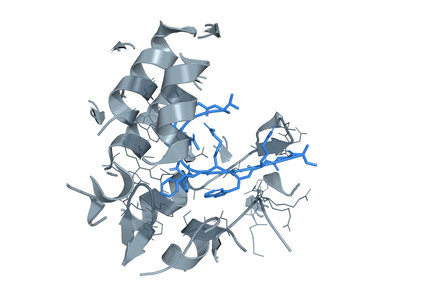
PyMOL render. Menin's pocket (grey; cartoon and lining side chains within 12 Å of the peptide) with KMT2A residues 4-15 (blue), the menin-binding motif every KMT2A fusion retains.
4GQ6 The interface the whole therapeutic programme is aimed at: 12 residues of KMT2A (MBM1) buried in menin's central cavity. This peptide is retained intact by the fusion.
Coordinates stream live from RCSB; nothing here is a drawing of a structure. Domain boundaries are the curated values in gene-kmt2a and protein-aff1-domains. Method and resolution for all eleven entries were read from the RCSB REST API rather than from memory. The junction itself has never been solved, and the residues either side of it are predicted to be disordered — which is the honest reason there is no picture of this protein.